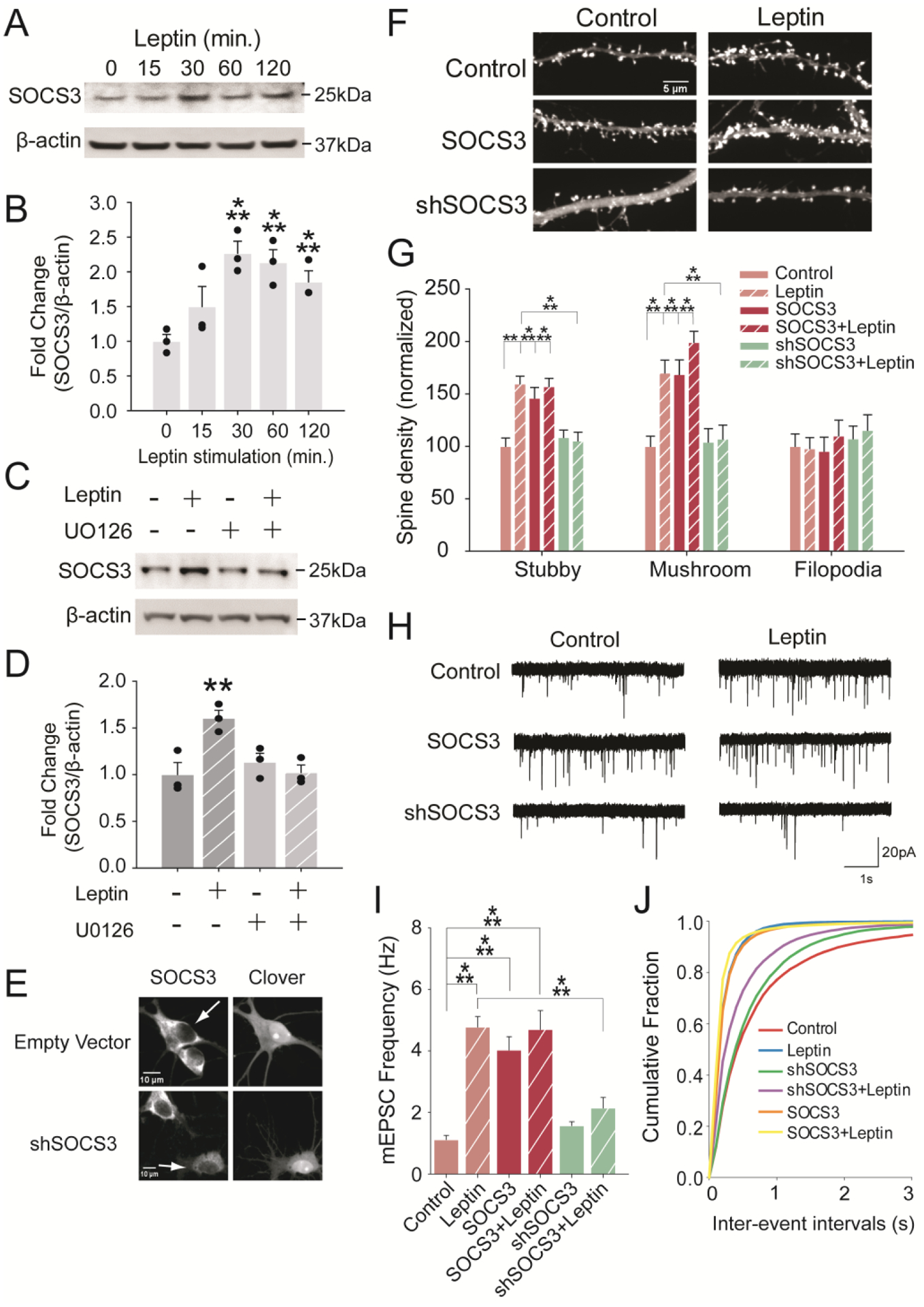
Figure 5:

SOCS3 increases functional spine formations in hippocampal neurons. (A, B) Leptin increased SOCS3 expression in DIV6–7 hippocampal neurons as early as 30 minutes of leptin stimulation. (C, D) Hippocampal neurons were stimulated with leptin (50nM) and/or U0126 (20μM) for 2 hours. Western blot was done with anti-SOCS3 and normalized against β-actin. All western blot data were analyzed with one-way ANOVA followed by Bonferroni multiple comparison. (E) Hippocampal neurons were transfected with pCAGGS-Clover ± shSOCS3 construct. Neurons were stained with anti-SOCS3. Transfected neurons were indicated with arrows. Note that in the upper left panel, there was no difference in SOCS3 levels between non-transfected and only pCAGGS-Clover (empty vector) transfected neurons. (F, G) DIV5–6 hippocampal neurons were transfected with Clover-βactin and either SOCS3 or shSOCS3, followed by leptin stimulation on DIV7. Neurons were fixed and analyzed on DIV12. (H, I) Hippocampal neurons were transfected and stimulated as in G, and mEPSCs were recorded. (J) Cumulative probability of the frequency of recorded mEPSC events. Both spine and electrophysiology data were analyzed with Kruskal-Wallis test followed by Dunn’s multiple comparison. Data were represented as mean ± SEM. Each dot represents one culture (***p<0.001, **p<0.01, *p<0.05).
