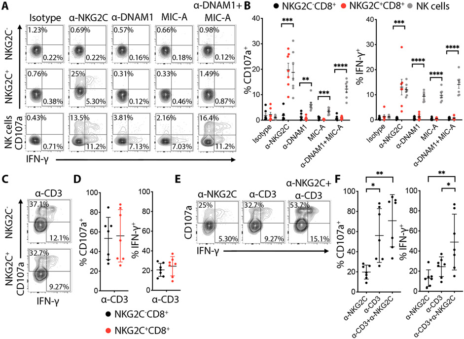
Fig. 6. NKG2C+CD8+ T cells express functional NKG2C that can cooperate with TCR activation in degranulation and IFN-γ production.
(A) Total PBMC cells were stimulated with the indicated plate-bound mAbs for 6 h. Representative flow cytometry plots showing degranulation (CD107a) and intracellular IFN-γ expression by pre-gated NKG2C+ or NKG2C−CD8+ T cells after triggering with plate-bound mAbs (10 μg/mL). (B) Graphs showing the percentage of CD107a+ and IFN-γ+ cells from 7 independent donors. Data are shown as mean ± SD. Statistical analysis was performed by t-test comparing the NKG2C−CD8+ vs NKG2C+CD8+ for each antibody with Holm-Sidak post-test correction. (C) FACS plots representing CD107a and intracellular IFN-γ expression by NKG2C− or NKG2C+CD8+ T cells after anti-CD3 (1 μg/mL) plate-bound stimulation. (D) Corresponding graphs showing the percentage of CD107a+ and IFN-γ+ cells from 7 independent donors. Data are shown as mean ± SD. (E) Representative FACS plots showing the percentage of CD107a+ cells after triggering of NKG2C+CD8+ T cells with plate-bound anti-CD3 or anti-NKG2C mAb alone or in combination. (F) Data are shown as the mean ± SD for 7 independent donors. Statistical significance was calculated by one-way ANOVA with Turkey’s multiple comparison test. * P-value ≤ 0.05, ** P-value < 0.01,***P-value ≤ 0.001, **** P-value < 0.0001.

