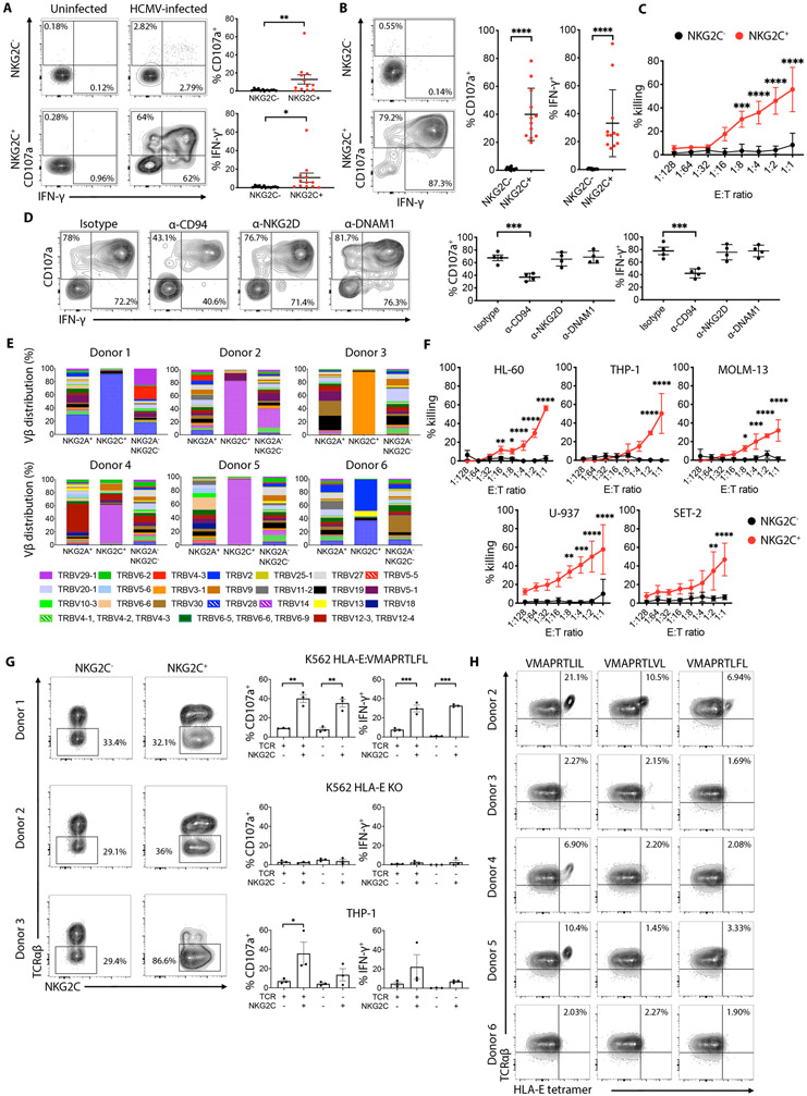
Fig. 7. NKG2C+CD8+ T cells anti-tumor and anti-HCMV effector functions are mediated by their NKG2C and TCR specificity.
(A) Representative flow cytometry plots showing degranulation (CD107a) and intracellular IFN-γ expression by pre-gated NKG2C+ or NKG2C−CD8+ T cells after 6 hours stimulation with uninfected or HCMV-infected human fibroblasts. Graphs on the right show cumulative analysis of CD107a+ and IFN-γ+ NKG2C+ or NKG2C−CD8+ T cells from 12 independent donors against uninfected or HCMV-infected human fibroblasts. Statistical significance was calculated using Wilcoxon matched-pairs signed rank test. (B) Representative flow cytometry plots showing degranulation (CD107a) and intracellular IFN-γ expression by pre-gated NKG2C+ or NKG2C−CD8+ T cells after 6 hours stimulation with K562 HLA-E:VMAPRTLFL target cells. Graph on the right show cumulative analysis of CD107a+ and IFN-γ+ NKG2C+ or NKG2C−CD8+ T cells from 12 independent donors against K562 HLA-E:VMAPRTLFL target cells. Statistical significance was calculated using Wilcoxon matched-pairs signed rank test. (C) In vitro cytotoxicity of FACS-sorted NKG2C+ or NKG2C−CD8+ T cells against K562 HLA-E:VMAPRTLFL target cells was assessed using a 6 hours bioluminescence assay. Results from 3 independent donors are shown. Statistic was calculated using a 2-way ANOVA comparing the mean of each effector to target (E:T) ratio between NKG2C− and NKG2C+CD8+ T cells. (D) Representative flow cytometry plots showing degranulation (CD107a) and intracellular IFN-γ expression by pre-gated NKG2C+CD8+ T cells against K562 HLA-E:VMAPRTLFL in the presence of the indicated blocking mAbs (10 μg/mL). Graphs on the right show cumulative analysis of 4 independent donors. Statistical significance was calculated using one-way ANOVA with Turkey’s multiple comparison test. (E) CD8+ T cell from 6 healthy donor PBMC were stained with a combination of different monoclonal antibodies against individual Vβ TCRs and analyzed by flow cytometry. Cells were pre-gated on NKG2A+NKG2C− (NKG2A+), NKG2A−NKG2C+ (NKG2C+) and NKG2A−NKG2C− CD8+ T cells. (F) In vitro cytotoxicity of FACS-sorted NKG2C+ or NKG2C−CD8+ T cells against the indicated AML target cell lines was assessed using a 6 hour bioluminescence assay. Results from 3 independent donors are shown. Statistic was calculated using a 2-way ANOVA comparing the mean of each E:T ratio between NKG2C− and NKG2C+CD8+ T cells (G) FACS plots showing TCRαβ expression on NKG2C+CD8+ or NKG2C−CD8+ T cells electroporated with Cas/9 and TRAC specific mRNA guides. Graphs on the right show cumulative analysis of CD107a+ and IFN-γ+ cells against the indicated targets. Statistical significance was calculated by one-way ANOVA with Turkey’s multiple comparison test. (H) NKG2C+CD8+ T cells from 5 donors were stained with the indicated HLA-E tetramers in the presence of a CD94 blocking antibody. ****P-value ≤ 0.0001, ***P-value ≤ 0.001, **P-value ≤ 0.01, *P-value ≤ 0.05.

