
Fig. 3.
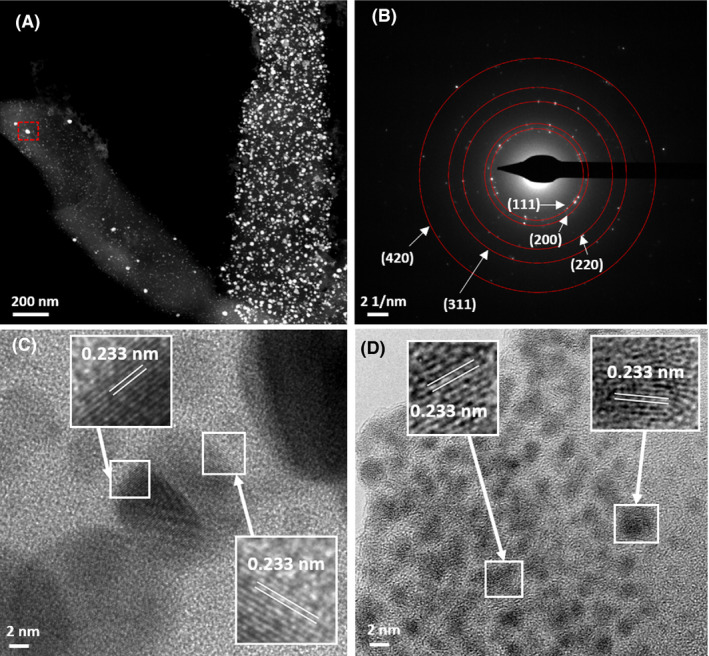
(A) STEM‐HAADF image of biosynthesized nanoparticles from Pd and Ag‐bearing solution and (B) corresponding polycrystalline select area diffraction pattern (from area highlighted in the red box) which is consistent with the FCC crystal structure of Ag(0). (C and D) Atomic resolution bright field images of small nanocrystals with corresponding lattice spacings consistent with the (111) facet of Pd(0).