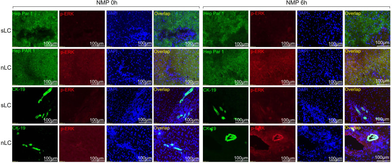
FIGURE 3.
Double immunofluorescence staining of p-ERK in livers with sLC and nLC at 0 h and 6 h NMP. The p-ERK signal is represented in red; the hepatocytes (labeled with hepatocyte specific antigen (Hep PAR-1) and cholangiocytes (labeled with CK-19) are represented in green; the nuclei are represented in blue (DAPI). CK-19, cytokeratin-19; DAPI, 4′,6-diamidino-2-phenylindole; p-ERK, phosphorylated extracellular signal-regulated kinase; nLC, nonsuccessful lactate clearance; NMP, normothermic machine perfusion; sLC, successful lactate clearance.

