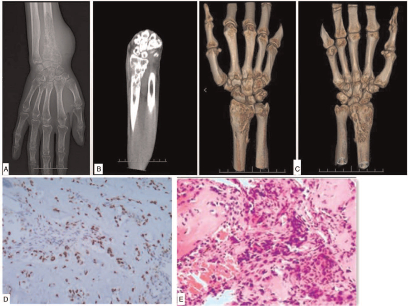
Figure 1.
Preoperative examination. (A–C) The x-ray film and computed tomography scan. multiple osteolytic bone destruction occurred at the distal end of the right radius and the surface of wrist joint, with several discontinuous bone cortex. The distal bone was slightly enlarged, with slight periosteal reaction. d.e pathological biopsy. (D) Preoperative biology and (E) second biology at the surgery: osteogenic malignant tumor.

