
Figure 1.
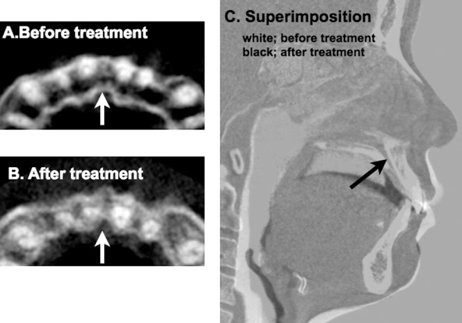
Cone beam computed tomography (CBCT) images of the incisive canal and superimposition before and after anterior retraction. Axial sections of the maxillary anterior region representing the apical one third of the maxillary incisors before (A) and after (B) anterior retraction. Notice the contact of the maxillary right central incisor and the approximation of the maxillary left central incisor roots to the incisive canal (arrow) after treatment. (C) CBCT superimposition on the cranial base before (in white) and after treatment (in black). Notice the changes in tooth position and the lateral profile following orthodontic treatment. No distinct changes were noted in the position of the incisive canal (arrow).