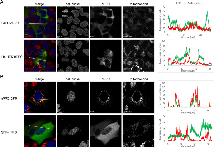
Fig 5. Localization of hPPO constructs in cell.
A; hPPO variants were transfected into in U-2 OS cells and visualized using PPOX-specific antibody (green channel) with a confocal microscope. Mitochondria were stained by Mitotracker Deep Red FM dye (red channel), whereas cell nuclei were visualized by DAPI staining (blue channel). B; Localization of GFP-tagged constructs was detected in live cells by a confocal microscope (green channel). Mitochondria were visualized by Mitotracker Deep Red FM (red channel), whereas cell nuclei were stained by Hoechst 33258 (blue channel). Charts on the right side show the colocalization of hPPO constructs and mitochondria marker Mitotracker in the area marked by a white line.

