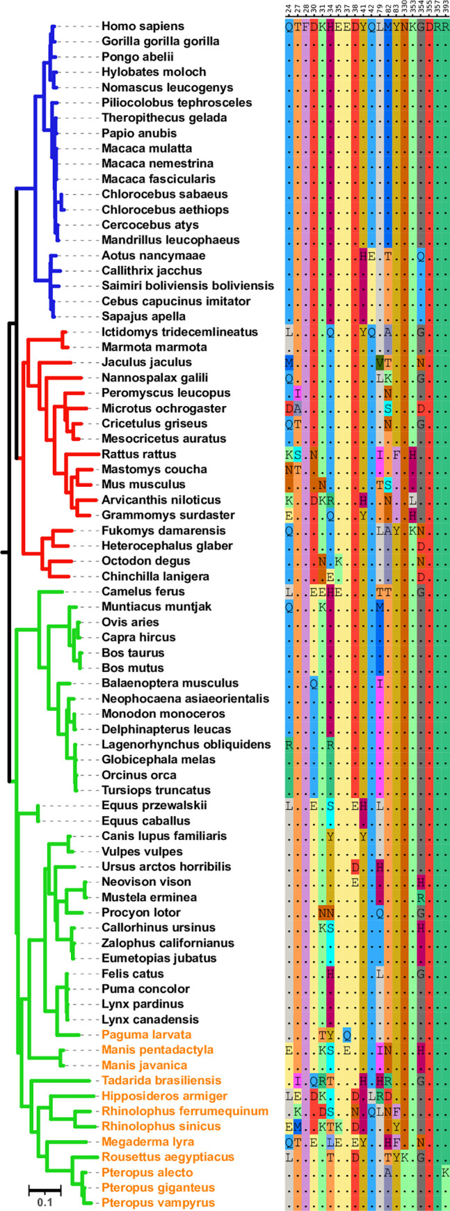
Fig 1. Phylogenetic tree of mammalian ACE2 sequences and residues that bind the viral spike protein.

(Left) A maximum likelihood phylogenetic tree for the ACE2 receptors with primates, rodent, and other mammal lineages shown in blue, red, and green, respectively. Bats, civet, and pangolin are specified via orange text. Only one species per genus is shown. The tree was built using RAxML [26] and the visualization was created using the iTOL platform [27]. (Right) For each of the 20 sites within the human ACE2 sequence that binds the SARS-CoV-2 spike protein, the corresponding residues in the multiple sequence alignment for the other ACE2 sequences are shown. Numbering is according to amino acid position in the human ACE2 sequence. The bat and rodent species clades exhibit the most amino acid variation.
