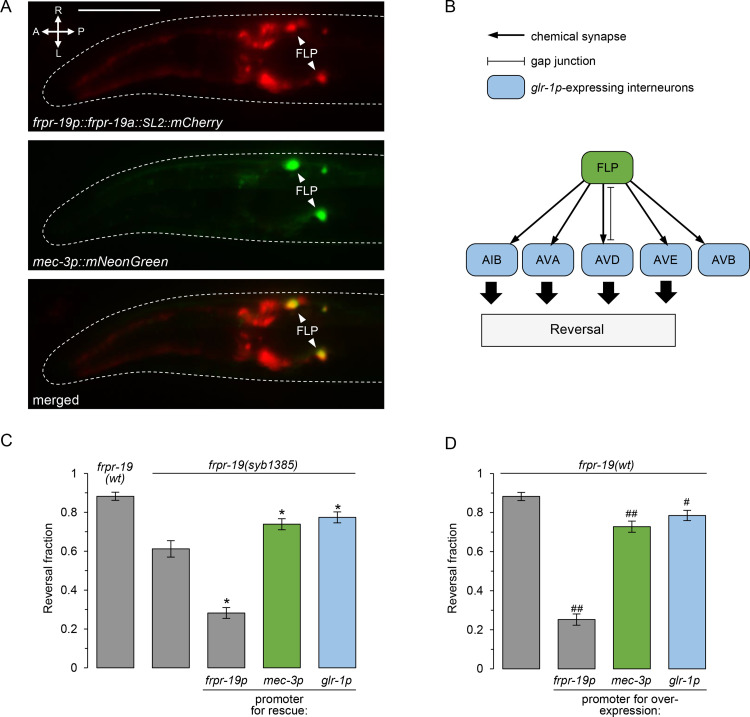
Fig 3. FRPR-19 acts cell autonomously in FLP neurons and downstream interneurons.
(A) Epifluorescence micrography of the head of a transgenic adult, showing the expression pattern of a transcriptional reporter for the frpr-19p promoter (mCherry, red, top), the localization of FLP with a mec-3p reporter (mNeonGreen, green, middle) and a merged picture (bottom). (B) Schematic showing FLP and all its postsynaptic partners who all express glr-1. (C) Cell-specific rescue experiments in the frpr-19(syb1385) null background showing a significant rescue effect with mec-3p promoter driving expression in FLP and with the glr-1 promoter driving expression in interneurons. Fraction of FLP optogenetic stimuli producing a reversal response, scored as in Fig 1. *, p < .01 versus frpr-19(syb1385) by Bonferroni contrasts. (D) Cell-specific overexpression experiments in frpr-19(wt) background showing a strong reversal decrease with the frpr-19p promoter and a partial decrease with mec-3p and glr-1p promoter. Fraction of FLP optogenetic stimuli producing a reversal response, scored as in Fig 1. ##, p < .01; #, p < .05 versus non-transgenic control by Bonferroni contrasts.

