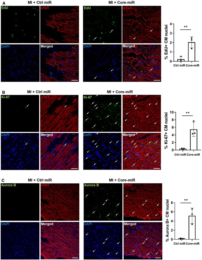
Fig. 6.
EV-derived-miR-106a–363 cluster activates cell cycle re-entry and proliferation in the injured myocardium. EdU incorporation of Control-miR and Core-miR injected mouse heart tissue was analyzed. EdU (50 μg/g) was injected via IP (intra-peritoneal) at day 3 post-MI. After 24 h, mice were sacrificed and their heart was collected. Cryo-sectioned tissue (8 μm/section) was stained by immunofluorescence specific for a EdU (green) and cTnT (red), b Ki-67 (green) and cTnT (red), and c Aurora B (green) and cTnT (red). Cells were counted from six random areas per each group (white arrows point to the double-positive cells). All quantitative graph showed the measurements of the number of cell cycle markers and cTnT double-positive cells in the peri-infarct region (PIR). Scale bar: 50 μm. Statistical significance was determined by unpaired two-tailed t-test and mean ± SD (n = 3). **P < 0.01

