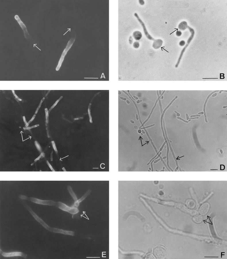
FIG. 1.
Phase-contrast (B, D, and F) and immunofluorescent (A, C, and E) photomicrographs of the same microscopic fields of C. albicans ATCC 66369 grown in medium 199 for 3 h at pH 6.7 (A and B) or 48 h (C and D) or C. albicans that originated from mouse abscesses (E and F) stained with MAb 16B1-F10. Note the homogeneous fluorescent labeling located solely on the filamentous cells; arrows point to the location of blastoconidia and cells that exhibited no fluorescence under UV illumination. Bars, 10 μm.

