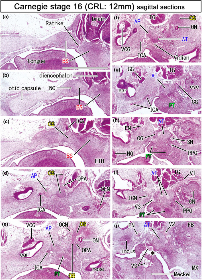
FIGURE 2.

Mesenchymal condensations for the initial sphenoid in embryos of 12 mm CRL. HE staining. Left‐hand side of each panel corresponds to the posterior site. Panel (a) displays the most medial site in the figure, while panel (J) the most lateral. The initial sphenoid was identified as five mesenchymal condensations: (1) the basisphenoid (BS; panels a–c) containing the Rathke's pouch (Rathke) and notochord (NC), (2) the orbitoshpenoid (OS) protruding superiorly (panels d and e), (3) the alar process (AP) near the developing inner ear (panels e and f) and, (4) the ala temporalis (AT), and (5) the pterygoid in the inferolateral side of the Vidian nerve (panels f and g). The sphenoid nerve (SN; panel h) transiently connects between the otic and pterygopalatine ganglia. All panels are prepared at the same magnification (scale bar in panel a, 1 mm). Other abbreviations, see the common abbreviation
