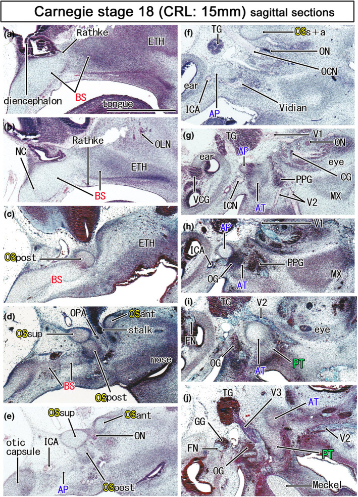
FIGURE 3.

Initial cartilages of the sphenoid in embryos of 15 mm CRL. Azan staining. Left‐hand side of each panel corresponds to the posterior site. Panel a displays the most medial site in the figure, while panel (j) the most lateral. The notochord is not contained in this figure. The orbitosphenoid (OS) is composed of the posterior, anterior and superior cartilage bars (OSpost, OSant, and OSsup) and they surround the optic nerve and ophthalmic artery (ON, OPA; panels d and e). The OSpost is attached to the basisphenoid (BS), while the OSsup and OSant joins in lateral sections (OSsup+ant in panel f). The pterygoid (PT) is still a mesenchymal condensation (panel j). All panels are prepared at the same magnification (scale bar in panel a, 1 mm). Other abbreviations, see the common abbreviation
