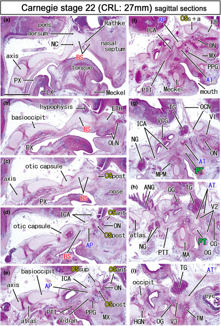
FIGURE 4.

Topographical anatomy of the skull base in an early fetus (27 mm). HE staining. Panel (a) displays the most medial site in the figure, while panel (i) the most lateral. Left‐hand side of each panel corresponds to the posterior site. Behind the hypophysis (panels a and b), a posterior protrusion of the basisphenoid (BS) is identified as the dorsum sellae (dorsum). Three cartilages of the OS (OSsup, OSpost, OSant) surrounds the optic nerve (ON; panel e). The oculomotor nerve (OCN) passes below the union of the OSsup and OSant of the OS (OSs+a; panel f). The maxillary nerve (V2) passes through the ala temporalis (AT; panels h). All panels are prepared at the same magnification (scale bar in panel a, 1 mm). Other abbreviations, see the common abbreviation
