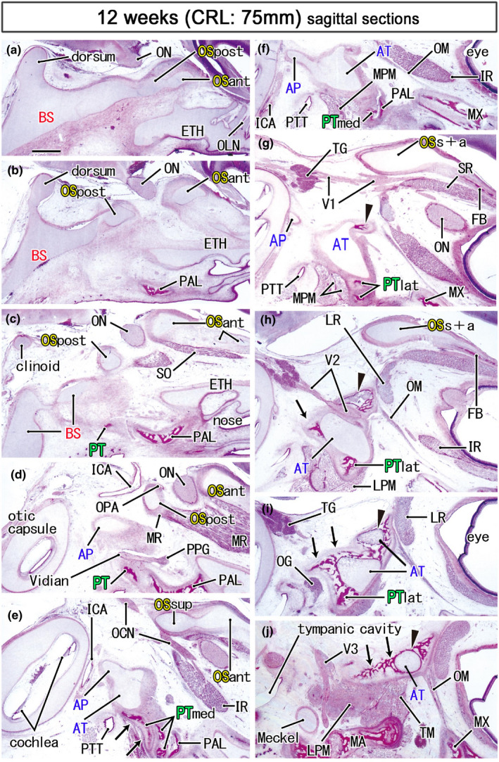
FIGURE 6.

Ossification in the sphenoid and nearby bones in a midterm fetus (75 mm). HE staining. Panel (a) displays the most medial site in the figure, while panel (j) the most lateral. Left‐hand side of each panel corresponds to the posterior site. The anterior part of the OS (OSant) extends anteriorly (panels a–e) and meets the bony frontal bone (FB; panels g and h). Ossification in the cartilaginous ala temporalis (arrowhead in panels g–j) makes a mosaic of bone and cartilage around the maxillary nerve (V2). Notably, the medial part of the pterygoid (PTmed) contains a cartilaginous bone (arrows in panel e). In the lateral side of the palatine bone (PAL), membranous bones in the lateral part of the pterygoid (PTlat; panels g and h) continue to the ala temporalis (arrows in panels h–j). The medial pterygoid muscle (MPM) attaches to not only the pterygoid but also to the ala temporalis (panels f and g). All panels are prepared at the same magnification (scale bar in panel a, 1 mm). Other abbreviations, see the common abbreviation
