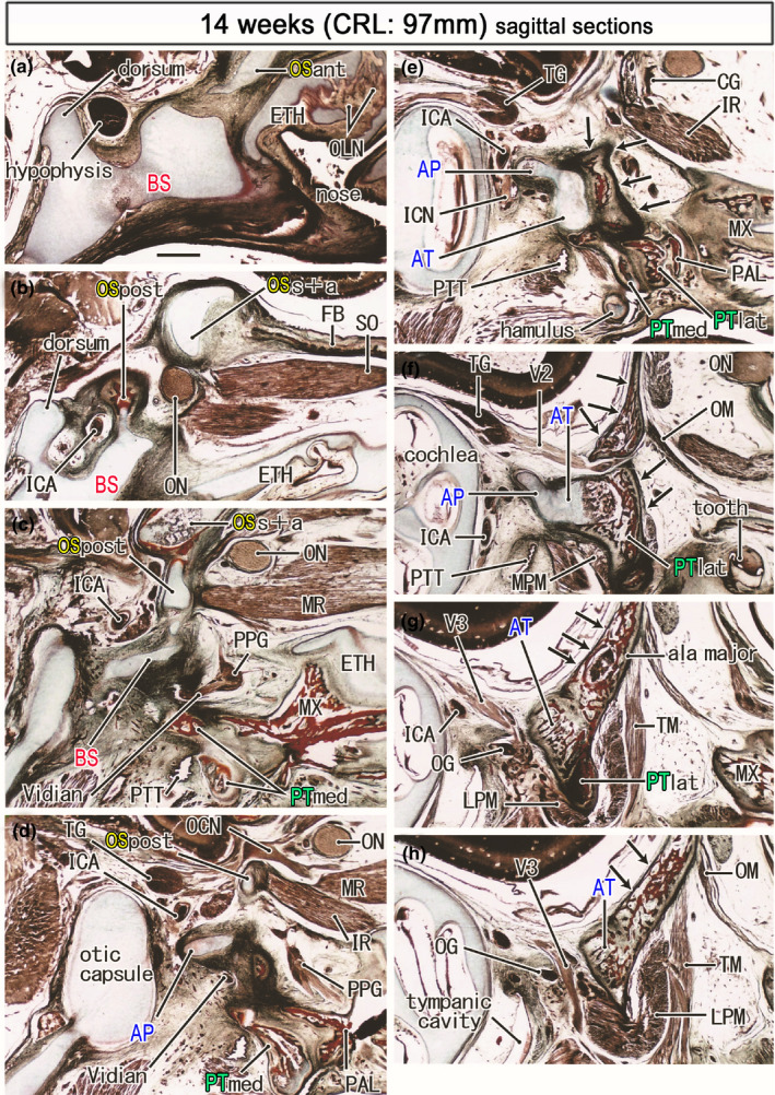
FIGURE 7.

Ossified pterygoid and the upper growth of the bony ala major in a midterm fetus (97 mm). Azan staining. Panel (a) displays the most medial site in the figure, while panel (h) the most lateral. Left‐hand side of each panel corresponds to the posterior site. The bony frontal bone extends posteriorly to meet the supero‐anterior part of the OS (OSs+a; panel b). The medial and inferior rectus muscles (MR, IR) originate from the posterior part of the OS (OSpost in panels c and d). The pterygoid (PTmed, PTlat) is ossified except for the hamulus (panel e). Membranous bones of the greater wing (ala major; arrows in panels e–h) extend along the posterior aspect of the ala temoralis and continue to lateral part of the pterygoid (panels f and g). All panels are prepared at the same magnification (scale bar in panel a, 1 mm). Other abbreviations, see the common abbreviation
