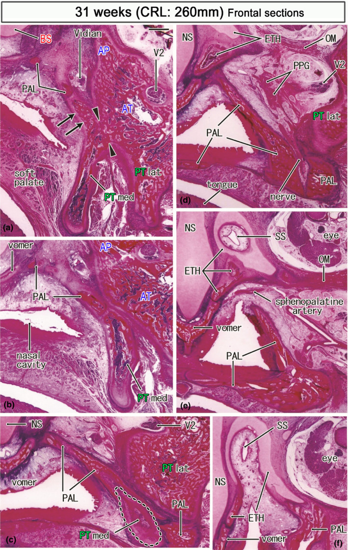
FIGURE 11.

Frontal sections of a specimen at 31 weeks (260 mm CRL). Panel (a) displays the most posterior section, while panel (f) the most anterior. Intervals between panels are 0.4 mm (a, b), 0.6 mm (b, c), 1.0 mm (c, d), 1.2 mm (d, e) and 1.0 mm (e, f). In panel (a), the medial pterygoid (PTmed) is fused with the lateral pterygoid (PTlat) at a site sandwiched by arrowheads. The former provides an upward protrusion (arrows in panel a). Posteriorly (panel d), the palatine bone (PAL) replaces the medial pterygoid (panels b and c) and approaches an ossified part of the ethmoid (ETH). The initial sphenoid sinus (SS) of this specimen is similar to that shown in Figure 9f: it is distant from the pterygoid and completely surrounded by the ethmoid (panels e and f). Panel (f) contains the future foramen sphenopalatinum through which the sphenopalatine artery runs to the nose. All panels are prepared at the same magnification (scale bar in panel a; 1 mm). Other abbreviations, see the common abbreviation
