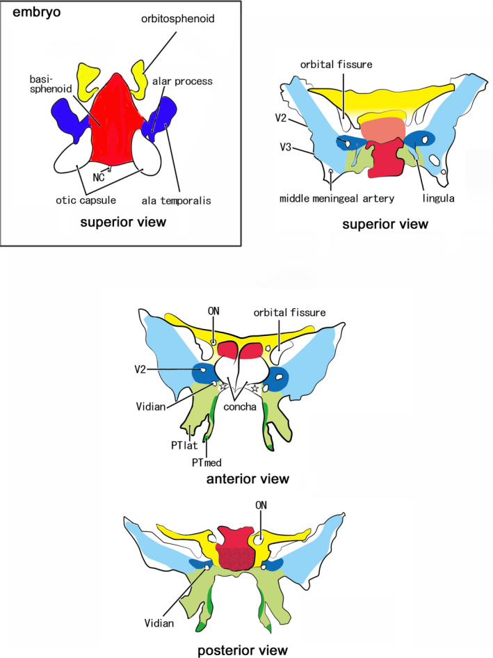
FIGURE 12.

Schematic represenations to connect fetal elements of the sphenoid to the adult morphology. Embryo: Red color displays the basisphenoid (BS). Blue color exhibits the alar process (AP) and the ala temporalis (AT). Yellow color indicates the orbitosphenoid (OS). Adult: Red color displays derivatives of the BS: a pale‐colored sella trucica in the superior view indicates the location deeper than the dorsum sellae. Blue color exhibits derivatives of the AT and AP: the pale blue indicates multiple membranous bones along and around the initial cartilages (dark blue). Green color shows the pterygoid: the limited dark green indicates endochondral bones for the hamulus and a marginal part of the medial pterygoid process (PTmed; anterior and posterior views). Yellow color indicates derivatives of the anterior, posterior, and superior bars of the OS. The OS makes not only the lesser wing but also a cartilage adjacent to the anterior margin of the BS (superior view). Star in the anterior view indicates a space occupied by the palatine bone and an ossified part of the ethmoid. ON, optic canal for the optic nerve; PTlat, lateral pterygoid process; V2, foramen rotundum for the maxillary nerve; V3, foramen ovale for the mandibular nerve; Vidian, sphenoid canal for the Vidian nerve
