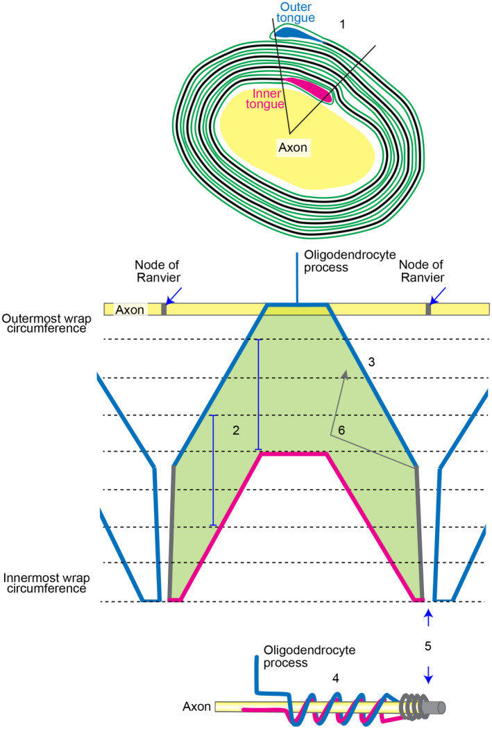
FIGURE 7.

Working model of the unwrapped myelin sheath. 1. Outer and inner tongues are located in the same quadrant in ~75% of fibres viewed in cross section (Peters, 1964). 2. The sheath (green; the shape shown here is simplified for clarity) extends only by growth at the inner tongue (Snaidero et al., 2014; magenta). Vertical blue lines indicate the distance the lamellipod has extended between the position where it first contacted the sheath (uppermost part) and the position where extension at the glial–axonal junction ceases (lowermost part). This distance is constant along the internode. 3. The lamellipod shape as it extends away from the oligodendrocyte process is fan‐shaped because the extreme leading edge (alone; eventually forming the inner tongue) drives protrusion forward and laterally from the point it hits the axon. 4. This fan shape gives rise to the corkscrew‐like spiralling (bell‐shaped and flat peaks) of the outer tongue as shown in our Figure 5. Thickening of the developing sheath proceeds (usually) from the middle of the internode (Figure 1 in Snaidero et al., 2014), but importantly, the mature sheath is equally thick along its length, thus the fan‐shaped outer aspect informs the shape of the inner aspect. (Not shown in the diagram—the lamellipod's shape is stochastic due to variation in the environment and random collisions; the distance extended must therefore be informed by the location of the outer tongue (blue) to achieve equal thickness along the internode). 5. The fan shape ends at the presumptive node, where the leading edge (growth zone) fails to spread any further laterally. The paranodal loops (grey) abut the node. 6. Secondary reverse growth from the growth zone, separate from the lamellipod and located on the outside of sheath, could contribute to the rings, bangles, plates and other conformations described by Río Hortega, and noted in the current work
