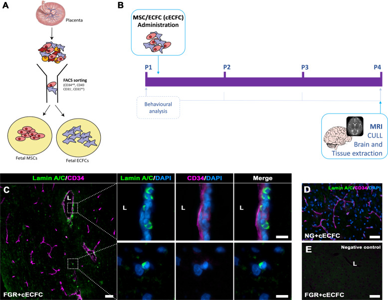
Fig. 1. Isolation and administration of combined Mesenchymal and Endothelial Colony Forming Cells.
A Foetal MSC and ECFCs were derived and isolated from healthy human placenta via FACs sorting. Isolated cells were cultured, enriched and prepared for intravenous administration as a combined stem cell preparation, termed cECFC (106 MSC and 106 ECFC). B Schematic representation of cECFC therapy in the newborn FGR pig model. C Presence of cells in treated FGR brain was confirmed post-mortem (P4) using Lamin A/C. Lamin A/C-positive cells were observed in the parenchyma as well as instances of vessel engraftment (lumen; L). D No Lamin A/C-positive labelling was observed in NG + cECFC tissue and E negative control (primary antibody omitted) (Scale bars: 50 µm; C high magnification: 10 µm).

