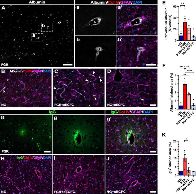
Fig. 3. cECFC administration ameliorates neurovascular integrity in the FGR neonate.
Representative labelling of endogenous albumin in the FGR as a marker of altered blood brain barrier integrity. A FGR brain showed albumin labelling (grey) predominantly localised to the perivascular space, between the lumen (L) and astrocyte endfeet (GFAP; magenta) (see Aa’-Ab’). B NG, C FGR + cECFC and D NG + cECFC displayed lower less overt albumin labelling. E A significantly higher percentage of vessels in FGR brain displayed perivascular labelling compared with NG. cECFC treatment did not significantly reduce the number of vessels with perivascular labelling. F FGR brain presented significantly greater albumin-positive labelling compared with NG. FGR + cECFC showed less labelling but was still elevated compared with NG. G Representative labelling of IgG (green) in the FGR brain showed extravasation into the brain parenchyma. Evident astrocyte activation (GFAP; magenta) was observed at vessels displaying altered permeability. H NG and J NG + cECFC displayed minimal IgG extravasation and maintained strong astrocyte interaction at the cerebrovasculature. I FGR + cECFC demonstrated less frequent IgG extravasation and significantly lower IgG-positive labelled area compared with untreated FGR K All values are expressed as mean ± SEM (minimum n = 6 for all groups). For K only brains pigs demonstrating IgG extravasation were included, NG (n = 1), FGR (n = 7), FGR + cECFC (n = 6), NG + cECFC (n = 1). Two-way ANOVA with Tukey post-hoc test (**p < 0.01, ****p < 0.001). For K; unpaired Student’s t test (*p < 0.05) (Scale bars: 50 µm; for A and G: low magnification: 200 µm).

