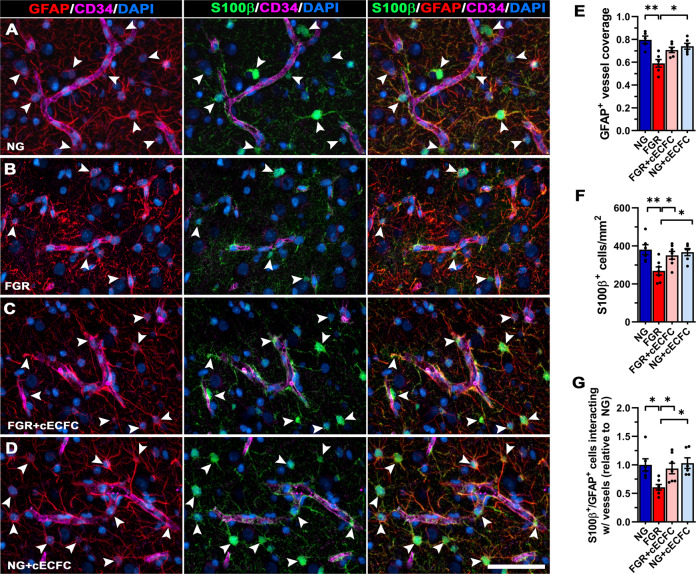
Fig. 4. Recovery of mature astrocytes at the NVU of FGR brain following cECFC administration.
Representative labelling for pan-astrocyte marker (GFAP; red) and mature astrocyte marker (S100β; green) in the cortex of pig at day 4. A NG showed strong S100β labelling co-localised to GFAP (arrowheads). These mature astrocytes demonstrate strong interaction of end-feet enveloping the length of neurovasculature. B FGR displayed intense thickened process labelling around the vasculature, indicative of reactive state. Less frequent S100β labelling was observed in the cortex as well as at the vasculature specifically. GFAP labelling along the vasculature was more sporadic and uneven when compared with NG. C FGR + cECFC displayed mature astrocytic labelling comparable to NG, with astrocyte end-feet displaying more contact with vasculature. D NG cECFC treated showed no alteration in cell count or morphology. E Co-localisation analysis demonstrated a decrease in GFAP-positive vessel coverage in FGR brain compared with NG. F The reduction in S100β cell counts in FGR was ameliorated following cECFC treatment. G Quantification of S100β-GFAP positive cells interacting with the vasculature confirmed a significant decrease in FGR relative to NG. FGR + cECFC displayed similar numbers of S100β-GFAP positive cells interacting with the vasculature to that observed in NG. All values are expressed as mean ± SEM (minimum n = 5 for all groups). Two-way ANOVA with Tukey post-hoc test (*p < 0.05) (Scale bars: 50 µm).

