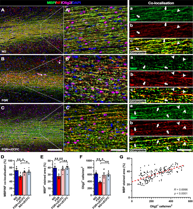
Fig. 7. Improved axonal myelination following cECFC treatment in FGR.
Representative labelling of myelin (MBP), neurofilament (NF) and pan-oligodendrocyte (Olig2) expression in white matter of NG, FGR and FGR + cECFC brains at postnatal day 4. A NG showed robust labelling for each marker with consistent presence of Olig2-positive cells along fibres. MBP and NF displayed strong co-localisation and structure throughout the white matter (A’a-A’c; arrows). B FGR display disrupted labelling of MBP and NF, with decreased Olig2-positive cells. There was observable disruption in labelling patterns with an evident loss in co-localisation of MBP to NF (B’; arrowheads). C FGR + cECFC brains showed normalised labelling and organisation of MBP and NF comparable that observed in NG. D Analysis of MBP/NF co-localisation in white matter shows a loss in axonal myelination in FGR compared with all groups. E Quantification of MBP-positive stained area (%) showed significantly decreased coverage and F decreased Olig2-positive cells/mm2 in FGR white matter relative to all groups, both of which were largely ameliorated following cECFC treatment. G Correlative analysis demonstrates a positive relationship between Olig2-positive cell count and MBP-positive stained area. All values are expressed as mean ± SEM (minimum n = 6 for all groups). Two-way ANOVA with Tukey post-hoc test (*p < 0.05, **p < 0.01, ***p < 0.001) (Scale bars: A–C: 200 µm; A’–C’ and a–c: 50 µm).

