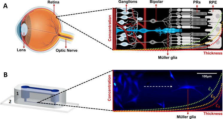
Fig. 1. Müller glia in vivo and in vitro.
A Schematic of the retina featuring Müller glia among other relevant cells of the retina, such as photoreceptors (PRs) and retinal pigmented epithelium (RPE) cells. This schematic features a representation of the vascular network within the retina. Schematics were created with BioRender.com. B rMC-1 model of Müller glia cells within a microchannel of the gLL microfluidic device (1: polymer, 2: glass slide) [13]. Cells were labelled with Calcein AM (2 μM). Concentration gradients are depicted in both the retina schematic and the microchannel of the gLL as a juxtaposition of the ability of both (in vitro and in vivo) platforms to develop concentration gradients ().

