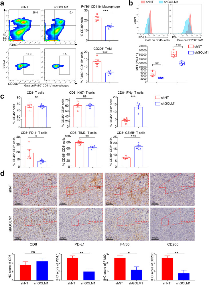
Fig. 2.
GOLM1 promotes TAM infiltration and PD-L1 expression on TAM in HCC. a Flow cytometric analysis of the infiltrated macrophages (F4/80+CD11b+) and TAMs (F4/80+CD11b+CD206+) in tumor tissues of subcutaneous implantation models of C57BL/6 mice with Hepa1-6-shNT or Hepa1-6-shGOLM1 cells (n = 5). b Flow cytometric analysis of PD-L1 expression in tumor and stromal cells (CD45−), and TAMs (F4/80+CD11b+CD206+) in tumor tissues of subcutaneous implantation models of C57BL/6 mice (n = 5). c Flow cytometry analysis of the number and functional state of CD8+ T cells in the subcutaneous implantation models of C57BL/6 mice. Inhibitory receptors (PD-1 and TIM-3), the ability to produce effector cytokines (IFN-γ and GZMB), and proliferation (Ki67) of CD8+ T cells were detected to evaluate the functional state of T cells (n = 5). d The densities of CD8, PD-L1, F4/80, and CD206 were consistently observed by IHC staining. The quantification is shown on the bottom panel (n = 5). *P < 0.05, **P < 0.01, ***P < 0.001, ns: no significant

