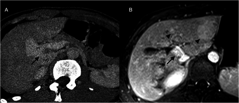
Fig. 1.
Type II congenital extrahepatic portosystemic shunt. A) CT mutiplanar reconstruction. A wide and short length shunt (black arrow) is seen connecting the inferior vena cava (IVC) and the portal trunk (PT). B) Axial equilibrium phase T1-weighted MR image with gadobutrol shows two slightly hyperintense nodules. These were also hyperintense on arterial and portal phases, compatible with the diagnosis of regenerative nodules (arrow heads)

