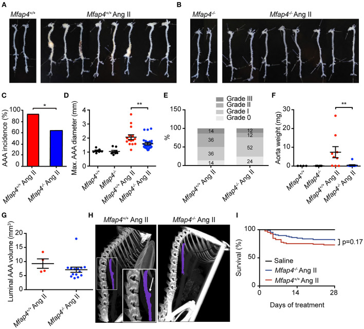
Figure 2.
Mfap4 deficiency attenuates Ang II-induced AAA development. (A,B) Representative photographs of dissected aortic segments from the aortic arch to the iliac bifurcation from (A) ApoE−/− (Mfap4+/+) and (B) ApoE−/−Mfap4−/− (Mfap4−/−) mice infused with saline (left panels) or Ang II (right panels) for 28 days. (C) AAA incidence. n = 15 (Mfap4+/+ Ang II), 26 (Mfap4−/− Ang II). (D) Quantification of maximal AAA diameter. n = 6–8 (saline), 15–26 (Ang II). (E) AAA severity scoring. n = 14 (Mfap4+/+ Ang II), 25 (Mfap4−/− Ang II). (F) Quantification of dissected aorta weight. n = 4–6 (saline), 9–12 (Ang II). (G) Quantification of luminal AAA volume assessed by micro-CT. n = 4 (Mfap4+/+ Ang II), 14 (Mfap4−/− Ang II). (H) Representative micro-CT images of the vascular luminal volume spanning a distance of 4 vertebrae after 28 days of Ang II infusion. Purple color demarks the investigated region of interest. The arrow indicates an aortic AAA. (I) Survival analysis. *p < 0.05, **p < 0.01, analyzed with Fisher's exact test (C) or Mann-Whitney U-test (D–F).

