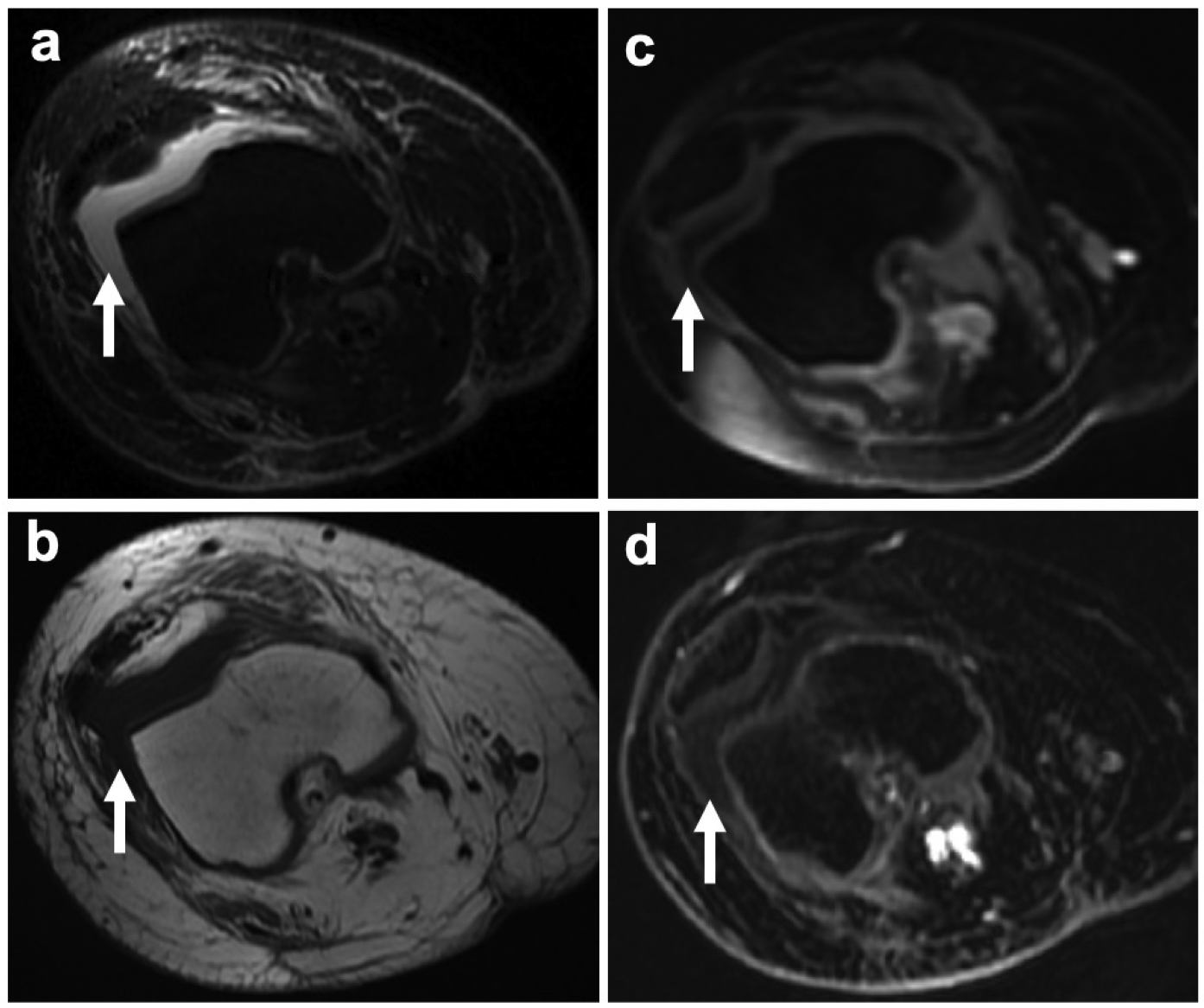
Fig. 4.

Knee joint effusion without T1-enhancement in a 24-year old female patient with recurrence of osteosarcoma in the right femur. a Axial PD-weighted FSE image shows a hyperintense knee joint effusion (arrow). b Corresponding pre-contrast axial T1-weighted FSE images demonstrates a hypointense joint effusion (arrow). c Post gadolinium and d 1 hour post ferumoxytol LAVA images show a hypointense knee joint effusion without T1-enhancement (arrows)
