
Figure 2. FAK regulates autophagy and AMPK signaling in adherent cells.
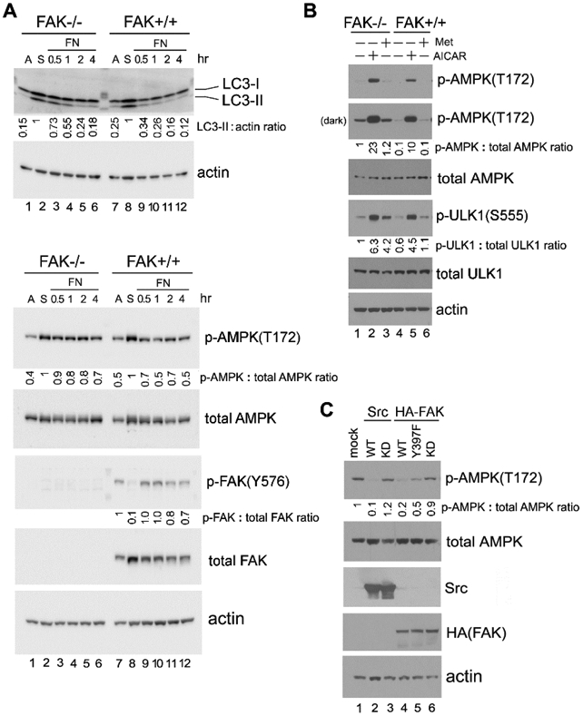
(A) “Detachment-re-attachment” experiment with FAK−/− and FAK+/+ MEFs. Adherent FAK−/− and FAK+/+ MEFs were either lysed for analysis (A = Adherent) or detached and kept in suspension for 1 hr. Suspended cells were then either lysed (S = Suspended) or replated onto FN-coated dishes for the indicated times, and immunoblot analysis was carried out with the indicated antibodies. Densitometric quantitation was performed and expressed as the fold change as in previous figures. Lane 2, suspended FAK−/− cells and lane 8, suspended FAK+/+ cells were used as the normalized control for lanes 1-6 and 7-12, respectively, in blots for LC3-II and p-AMPK(T172). Lane 7, adherent FAK+/+ cells was used as the normalized control for the p-FAK(Y576) blot. In all panels for Fig. 2, results shown are representative of at least three independent experiments with similar results. (B) Activation of AMPK in FAK−/− and FAK+/+ MEFs. Cells were treated (+) or not (−) with the AMPK agonists AICAR (2 mM) or Metformin (Met; 2 mM) for 1 hr, followed by immunoblot analysis and quantification as above (lane 1, untreated FAK −/− cells was used as the normalized control). “Dark” indicates 5 times longer exposure compared to the top panel. (C) Effect of Src or FAK overexpression on AMPK-T172 phosphorylation. 293T cells were transfected or not (mock) to express the various Src and FAK proteins as indicated (WT=wild-type; KD=kinase-dead). Total cell lysates were then subjected to immunoblot analysis with the indicated antibodies. Quantification was performed as above using mock-transfected cells (lane 1) as the normalized control. For quantification and statistical analysis of the independent blots for Figs. 2A, B and C, please see Supplementary Figure 1.