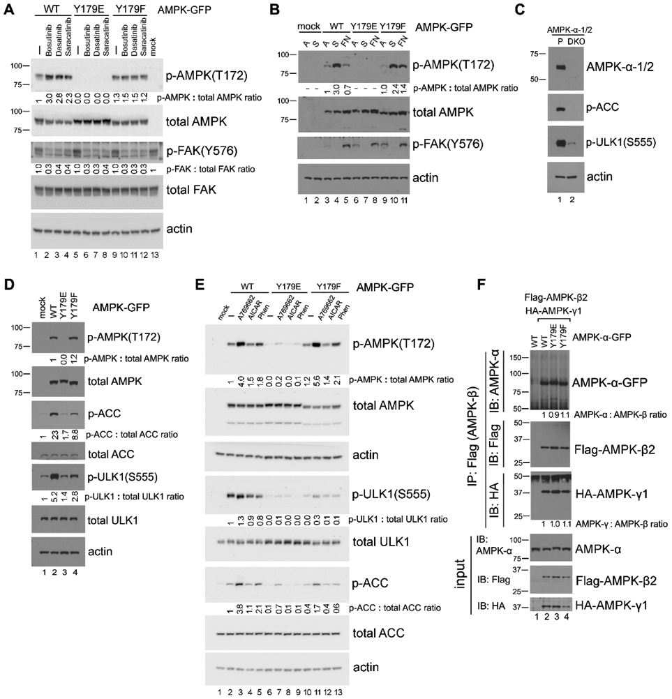
Figure 5. The AMPK-Y179 site plays a role in regulating phosphorylation of AMPK at T172.

(A) Effect of Src inhibition on AMPK-T172 phosphorylation. 293T cells transfected with the various GFP-tagged AMPK-α2 constructs were treated or not (−) with 10 μM of Src kinase inhibitors Bosutinib, Dasatinib or Saracatinib for 1 hr, and AMPK-α2-GFP phosphorylation on T172 was detected by immunoblot in total cell lysates. Quantification was performed by using untreated WT-AMPK cells (lane 1) as the normalized control. In all panels for Fig. 5, results shown are representative of at least three independent experiments with similar results. (B) “Detachment-re-attachment” experiment. 293T cells transfected with the various GFP-tagged AMPK-α2 constructs were lysed when adherent (A) or kept in suspension (S) for 1 hr before replating of the cells to FN-coated dishes for 2 hr. AMPK-α2-GFP phosphorylation on T172 was detected by immunoblot in total cell lysates as above. Quantification was performed by using adherent WT-AMPK cells (lane 3) as the normalized control. (C) Immunoblot analysis of parental 293A (P) and 293A-AMPK1/2-DKO cells with the indicated antibodies. (D) Expression of various AMPK-Y179 mutant proteins in 293A-AMPK1/2-DKO cells. Cells were transfected with the various GFP-tagged AMPK-α2 constructs, and immunoblot analysis was carried out with the indicated antibodies. Quantification was performed with the WT-AMPK cells (lane 2) as the normalized control for the p-AMPK(T172) blot, and with mock-transfected cells (lane 1) as the normalized control for the p-ACC and p-ULK1(S555) blots. (E) Activation of AMPK in 293A-AMPK1/2-DKO cells transfected with the various GFP-tagged AMPK-α2 constructs. Cells were treated or not (−) with various AMPK agonists (200 μM A-769662, 2 mM AICAR or 1 mM phenformin, as indicated) for 1 hr, followed by immunoblot analysis as above. Fold change of p-AMPK-T172, p-ULK1-S555, and p-ACC was quantified, normalized to that of the total corresponding protein and compared to untreated WT-AMPK-transfected cells (lane 2). (F) Examination of AMPK subunits assembly. Co-IP was performed in lysates from 293T cells that had been co-transfected with Flag-tagged AMPK-β2 and HA-tagged AMPK-γ1, along with GFP-tagged AMPK-α constructs by using anti-Flag antibody, and the associated AMPK subunits were detected by immunoblot with the relevant antibodies. Quantification was performed with WT-AMPK cells (lane 2) as the normalized control. For quantification and statistical analysis of the independent blots for Figs. 5A, B and E, please see Supplementary Figure 1.
