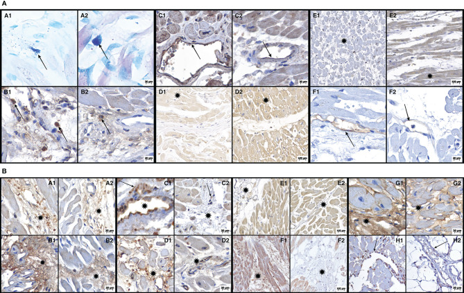
Figure 1.
(A) Photomicrographs demonstrating histochemical (A1/2 - Toluidine Blue) and immunohistochemical (B = CD163; C = Casp-1; D = Casp-9; E = GSDM-D; F = ICAM-1) reactions of both groups: cases of COVID-19 (B1-F1) and cases of their respective controls (B2-F2). A1 (COVID-19) and A2 (control) show a mast cell in the perivascular space (arrows): in A1 the mast cell is degranulated, whereas in A2, the mast cell is intact. B1 (COVID-19) shows CD163-immunostained macrophages (arrows) in the myocardial interstitium, whereas in B2 (control) these CD163 macrophages (arrow) are less numerous. C1 (COVID-19) shows endothelial cells (arrow) with strong expression of Casp-1, whereas in C2 (control), Casp-1 expression in endothelial cells (arrow) is much more discrete. D1 (COVID-19) shows a weak expression of Casp-9 in the myocardium (asterisk), whereas in D2 (control), this expression is strong (asterisk). E1 (COVID-19) shows a weak expression of GSDM-D in the myocardium (asterisk), with E2 (control) showing a strong expression (asterisk). F1 (COVID-19) shows a strong expression of ICAM-1 in endothelial cells (arrow), whereas, in F2 (control), this expression is discrete (arrow). (B) Photomicrographs demonstrating immunohistochemical reactions (A = TNF-α; B = MMP-9; C = IL-4; D = IL-6; E = IL-1β; F = TGF-β; G = collagen 3; H = TUNEL) from both groups: cases of COVID-19 (A1-H1) and their respective controls (A2-H2). A1 (COVID-19) shows myocardial interstitium expressing TNF-α (asterisk), whereas, in A2 (control), we can observe that this expression is discrete (asterisk). B1(COVID-19) shows myocardial interstitium expressing MMP-9 (asterisk), whereas, in B2 (control), this expression is discrete (asterisk). C1 (COVID-19) shows interstitial macrophages (arrow) and endothelial cells (asterisk) with strong expression of IL-4, whereas in C2 (control), the expression of IL-4 in interstitial macrophages (arrow) and endothelial cells (asterisk) is discrete. D1 (COVID-19) shows a strong expression of IL-6 in the interstitium (asterisk), whereas, in D2 (control), this expression is discrete (asterisk). E1 (COVID-19) shows moderate expression of IL-1β in the myocardial interstitium (asterisk), and E2 (control) has a more discrete expression (asterisk). F1 (COVID-19) shows a strong expression of TGF-β in the interstitium of the myocardium (asterisk), whereas, in F2 (control), this expression is much more discrete or absent (asterisk). G1 (COVID-19) shows a strong expression of collagen 3 in the interstitium of the myocardium (asterisk), and this expression is much more delicate (asterisk) in G2 (control). H1 (COVID-19) demonstrates elevated DNA fragmentation in the TUNEL assay (arrow) compared to H2 (control), where few nuclei (arrow) are labeled.

