Fig. 1.
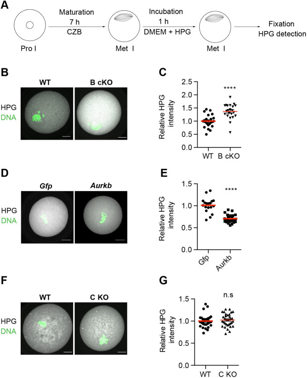
Translation is upregulated in AURKB cKO oocytes. (A) Schematic of HPG assay. (B-G) Metaphase I (Met I) oocytes were labeled with HPG and stained with anti-HPG (gray) and DAPI (DNA, green). (B) WT and AURKB cKO (B cKO) mice. (C) Relative intensity of HPG from B. Values normalized to WT (number of oocytes: WT 24, B cKO 22). (D) B cKO oocytes microinjected with Gfp or Aurkb cRNA. (E) Values normalized to Gfp-injected (number of oocytes: Gfp 18, Aurkb 24). (F) Met I oocytes from WT and AURKC KO (C KO). (G) Relative pixel intensity of HPG from F. Values normalized to WT (number of oocytes: WT 29, C KO 33). n.s., not-significant (P=0.4240). Experiments replicated three times; one mouse/genotype/replicate. Data points show individual oocytes. Red horizontal line indicates the mean. ****P<0.0001 (unpaired two-tailed Student's t-test). Scale bars: 10 µm.

