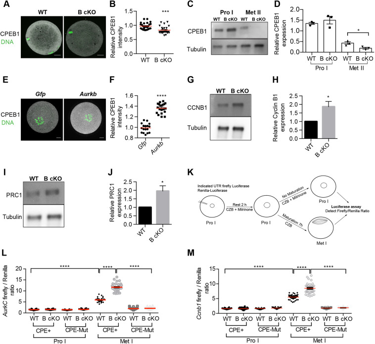
Fig. 2.
CPEB1 activity is increased in AURKB cKO oocytes. (A) Met II eggs from WT and AURKB cKO (B cKO) mice stained with anti-CPEB1 (gray) and DAPI (DNA, green). (B) Relative intensity of CPEB1 from A. Values normalized to WT (number of oocytes: WT 27, B cKO 25). (C) Representative western blot detecting CPEB1 from WT and B cKO (30/lane). Loading control: α-tubulin. (D) Quantification of CPEB1 after normalizing C to α-Tubulin in three experiments. (E) B cKO oocytes were microinjected with Gfp or Aurkb cRNA, matured to Met I and stained with anti-CPEB1 (gray) and DAPI (DNA, green). (F) Relative CPEB1 intensity from E. Values normalized Gfp-injected (Number of oocytes: Gfp-17; Aurkb-20). (G,I) Western blot of Met I oocytes detecting CCNB1 (25/lane; G) or PRC1 (25/lane; I). (H,J) Relative CCNB1 or PRC1expression from G and I, respectively. Values normalized to α-tubulin. (K) Schematic of luciferase assay. (L,M) Prophase I (Pro I) oocytes were co-injected with luciferase RNAs: Aurkc (L) and Ccnb1 (M) fused with CPE+ or CPE-mutated (Mut) UTR. Luminescence was measured and quantified as firefly/Renilla (number of oocytes: L – WT-CPE+ 29, WT-CPE-Mut 30, B cKO-CPE+ 29, B cKO-CPE-Mut 27; M – WT-CPE+ 40, WT-CPE-Mut 34, B cKO-CPE+ 39, B cKO-CPE-Mut 33). Experiments repeated three times; total 3-5 mice/genotype. Red horizontal line indicates the mean. *P<0.05; ***P<0.001; ****P<0.0001 (unpaired two tailed Student's t-test). Scale bars: 10 µm.

