Fig. 3.
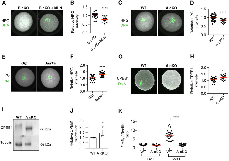
AURKA is required for translation and CPEB1 regulation. (A) Metaphase I (Met I) oocytes from AURKB cKO (B cKO) treated with DMSO or 1 µM MLN8237 (MLN), labeled with HPG and stained with anti-HPG (gray) and DAPI (DAPI, green). (B) Relative intensity of HPG from A (number of oocytes: B cKO 23; B cKO+MLN 27). (C) Met I oocytes from WT and AURKA cKO (A cKO) mice were labeled with HPG and stained with anti-HPG (gray) and DAPI (DNA, green). Shown are representative confocal z-projections. (D) Relative intensity of HPG from C. Values normalized to WT (number of oocytes: WT 39, A cKO 39). (E) A cKO oocytes were microinjected with Gfp or Aurka cRNA, matured to Met I, labeled with HPG and stained with anti-HPG (gray) and DNA (DAPI, green). (F) Values normalized to Gfp-injected group (number of oocytes: Gfp 18, Aurka 21). (G) Met I oocytes from WT and A cKO mice were stained with anti-CPEB1 (gray) and DAPI (DNA, green). (H) Relative intensity of CPEB1 from G. Values normalized to WT (number of oocytes: WT 27, A cKO 26). (I) Met I oocytes from WT and A cKO mice were collected for western blotting to detect CPEB1 (30/lane). Loading control: α-tubulin. (J) Quantification of CPEB1 after normalizing I to α-tubulin. (K) Prophase I (Pro I) oocytes were co-injected with luciferase RNAs as in Fig. 2M. Luminescence was measured and quantified (number of oocytes: WT 36, A cKO 32). Experiments A-F replicated three times; one mouse/genotype/replicate; experiments G-I repeated three times; total 3-5 mice/genotype. Red horizontal line indicates the mean. *P<0.05; **P<0.01; ****P<0.0001 (unpaired two tailed Student's t-test). Scale bars: 10 µm.

