Fig. 4.
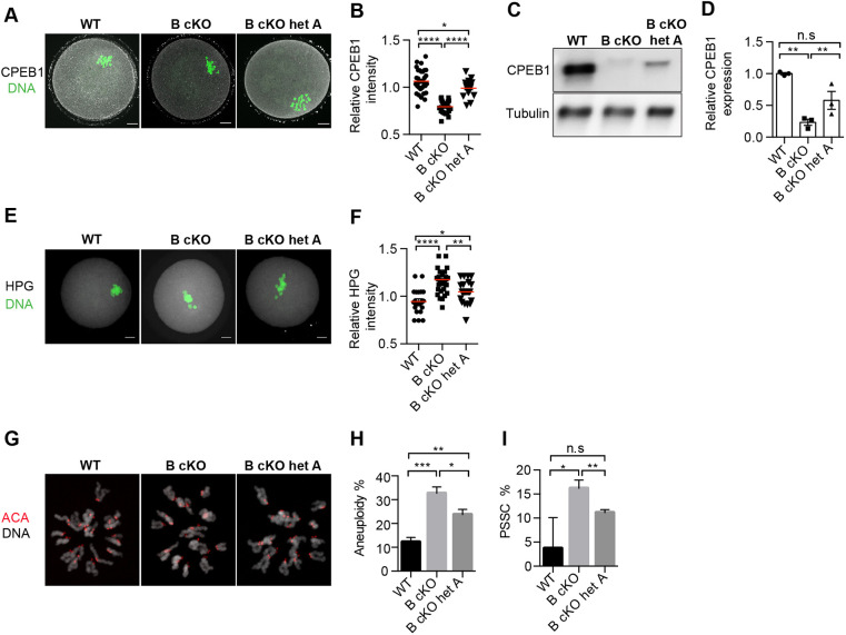
Reduction of AURKA activity in AURKB cKO oocytes partially rescues CPEB1, translation and meiosis defects. (A) Metaphase I (Met I) oocytes from WT, AURKB cKO (B cKO) and AURKB cKOs heterozygous for Aurka (B cKO het A) mice were stained to detect CPEB1 (gray) and DAPI (DNA, green). (B) Relative intensity of CPEB1 from A. Values normalized to WT (number of oocytes: WT 34, B cKO 25, B cKO het A 24). (C) Met I oocytes from WT, B cKO and B cKO het A mice were collected for western blotting to detect CPEB1 (30/lane). Loading control: α-tubulin. (D) Quantification of CPEB1 after normalizing C to α-tubulin. (E) Met I oocytes were stained to detect HPG (gray) and DAPI (DNA, green). (F) Relative intensity of CPEB1 from E. Values normalized to WT (number of oocytes: WT 22, B cKO 30, B cKO het A 21). (G) Representative images of chromosome spreads of Metaphase II eggs stained with Anti-Centromeric Antibodies (ACA, red) and DAPI (gray). (H) Quantification of the aneuploidy. (I) Quantification of the premature separation of sister chromatids (PSSC). Number of oocytes in H,I: WT 29, B cKO 31, B cKO het A 36. Experiments repeated three times; total 3-4 mice/genotype. Red horizontal line indicates the mean. Error bars indicate s.e.m. *P<0.05; **P<0.01; ***P<0.001; ****P<0.0001 (one-way ANOVA). n.s., not significant. Scale bars: 10 µm.

