Fig. 6.
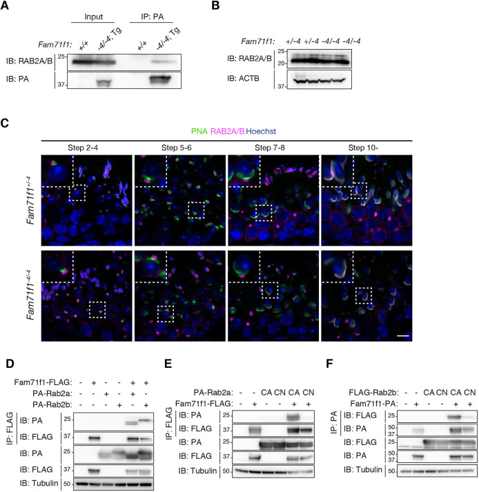
FAM71F1 interacts with the constitutively active form of RAB2A/B. (A) Immunoprecipitation of FAM71F1-PA using anti-PA antibodies. Testis lysates of wild-type (+/+) or Fam71f1-PA Tg (−4/−4; Tg) mice were used. FAM71F1-PA interacts with RAB2A/B. (B) Immunoblotting analysis for RAB2A/B using testis lysates. The amounts of RAB2A/B were comparable between Fam71f1+/−4 and Fam71f1−4/−4 mice. ACTB was used as a loading control. (C) Immunofluorescence staining of RAB2A/B and the acrosome during spermatogenesis using testis sections. RAB2A/B was localized in the Golgi apparatus until steps 7 to 8 and in the acrosome around step 10. No differences were found in the RAB2A/B localization between Fam71f1+/−4 and Fam71f1−4/−4 mice. Higher magnification images of the boxed areas are shown in the top-left. Nuclei are stained blue (Hoechst), acrosomes green (lectin-PNA) and RAB2A/B magenta. (D) Immunoprecipitation of FAM71F1-FLAG using anti-FLAG antibodies. Fam71f1-FLAG was transiently expressed with PA-Rab2a or PA-Rab2b in HEK293T cells. FAM71F1-FLAG interacts with both PA-RAB2A and PA-RAB2B. (E) Immunoprecipitation of FAM71F1-FLAG using anti-FLAG antibodies. PA-Rab2a (CA or CN) and Fam71f1-FLAG were transiently expressed in HEK293T cells. FAM71F1 interacts with RAB2A (CA). (F) Immunoprecipitation of FAM71F1-PA using anti-PA antibodies. FLAG-Rab2b (CA or CN) and Fam71f1-PA were transiently expressed in HEK293T cells. FAM71F1 interacts with RAB2B (CA). Tubulin was used as a loading control in D-F. Scale bar: 10 μm in C. CA, constitutively active (Q65L); CN, constitutively negative (S20N).

