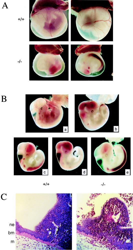
FIG. 2.
Phenotype of Fli1 mutant embryos. (A) Absence of blood in the yolk sac of Fli1 mutant E11 embryos. Representative photographs of E11 embryos with intact visceral yolk sacs are shown. Blood-filled vessels in the yolk sac are seen in the wild-type littermate (+/+) but not in the homozygous mutant (−/−). (B) Fli1 mutant E11 embryos hemorrhage. E11 homozygous mutant embryos dissected free of the yolk sac are easily distinguished from their wild-type littermates (a) by their smaller overall size and hemorrhaging of fetal red cells into the cephalic ventricles (b to d) and the central canal of the neural tube (b, c, and e). (C) Histological analysis of E11 homozygous mutant embryos. Transverse sections through E11 embryos demonstrate a disruption in the columnar neuroepithelium, reduced adjacent extracellular matrix, and a hematoma at the site of hemorrhage (right panel). A section from a wild-type embryo is shown on the left for comparison. Note the nucleated red cells in the lumen of the neural tube (top, right panel). Positions of neuroepithelial (ne) and mesenchymal (m) cells and basement membrane (bm) are indicated.

