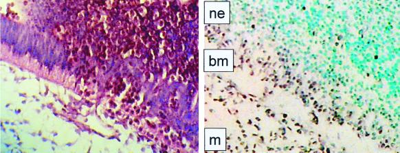
FIG. 3.
Expression of Fli1. In situ hybridization with antisense Fli1 RNA of transverse sections of wild-type E10 embryos was performed. Embryo sections (8 μm) were processed for in situ hybridization and counterstained with methyl green. Positions of neuroepithelial (ne) and mesenchymal (m) cells and basement membrane (bm) are indicated in the in situ-hybridized section (right panel). A comparable, hematoxylin-and-eosin-stained section from the same region of a mutant E11 embryo is shown for comparison (left panel), demonstrating disruption of the thick basement membrane and columnar neuroepithelium and pockets of nucleated red cells within these tissues.

