Figure 3.
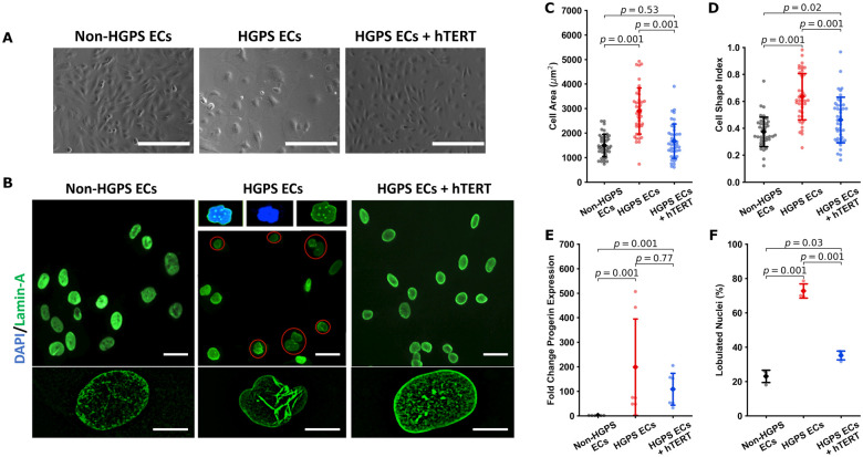
hTERT treatment normalizes cellular and nuclear morphology in Hutchinson-Gilford progeria syndrome endothelial cells. Representative bright field imaging of non-Hutchinson-Gilford progeria syndrome, Hutchinson-Gilford progeria syndrome, and hTERT treated Hutchinson-Gilford progeria syndrome endothelial cells are shown in (A) (scale bar—200 µm). Nuclear lamin A staining is shown in (B) (scale bar: top—30 µm; bottom—10 µm). Hutchinson-Gilford progeria syndrome endothelial cells show a compromised morphology, having increased cellular area (n = 43 cells across three replicates, C) and a more rounded morphology as indicated by an increased cell shape index/circularity index (n = 43 cells across three replicates, D). Cell shape index/circularity is defined as 4π(area/perimeter2). hTERT therapy additionally induced a downward trend in progerin expression as assessed via RTqPCR (n = 6) (E) and improves nuclear morphology (F), reducing the number of folded nuclei (three experimental replicates, percent folded nuclei reported for at least 300 cells in each group per replicate). Data were quantified using a one-way ANOVA with post hoc Tukey’s test (ddCt analysis for qPCR), significance taken at P < 0.05. All between-group statistics are displayed natively. All error bars show standard deviation.

