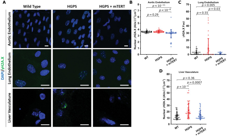
Figure 8.
mTERT Treatment in Hutchinson-Gilford progeria syndrome mice reduces DNA damage markers in aorta, lung and liver endothelial cells. Representative images from aortic endothelial cells, lung endothelial cells, and liver vasculature are displayed in (A) (scale bar—10 µm). All tissues were stained for nuclei (DAPI, blue) and γH2A.X (green). Individual cells from the tissues were quantified (n > 50) for nuclear γH2A.X fluorescence (tissue sections from aorta in B and liver in D) and γH2A.X foci in isolated mouse lung endothelial cells in (C). In the aortic and lung tissue, differences were not statistically significant, though γH2A.X was significantly downregulated or decreased in the mTERT treatment groups. In liver tissues, Hutchinson-Gilford progeria syndrome vascular cells displayed increased γH2A.X, which was reversed by mTERT. All data collected from four to five different mice histology sections for each group. The data were tested for normality using D’Agostino-Pearson test. Kruskal–Wallis with post hoc Dunn test was used to quantify the data. Significance was taken at P < 0.05, all between-group statistics are displayed natively. All error bars show standard deviation. DAPI, 4′,6-diamidino-2-phenylindole.

