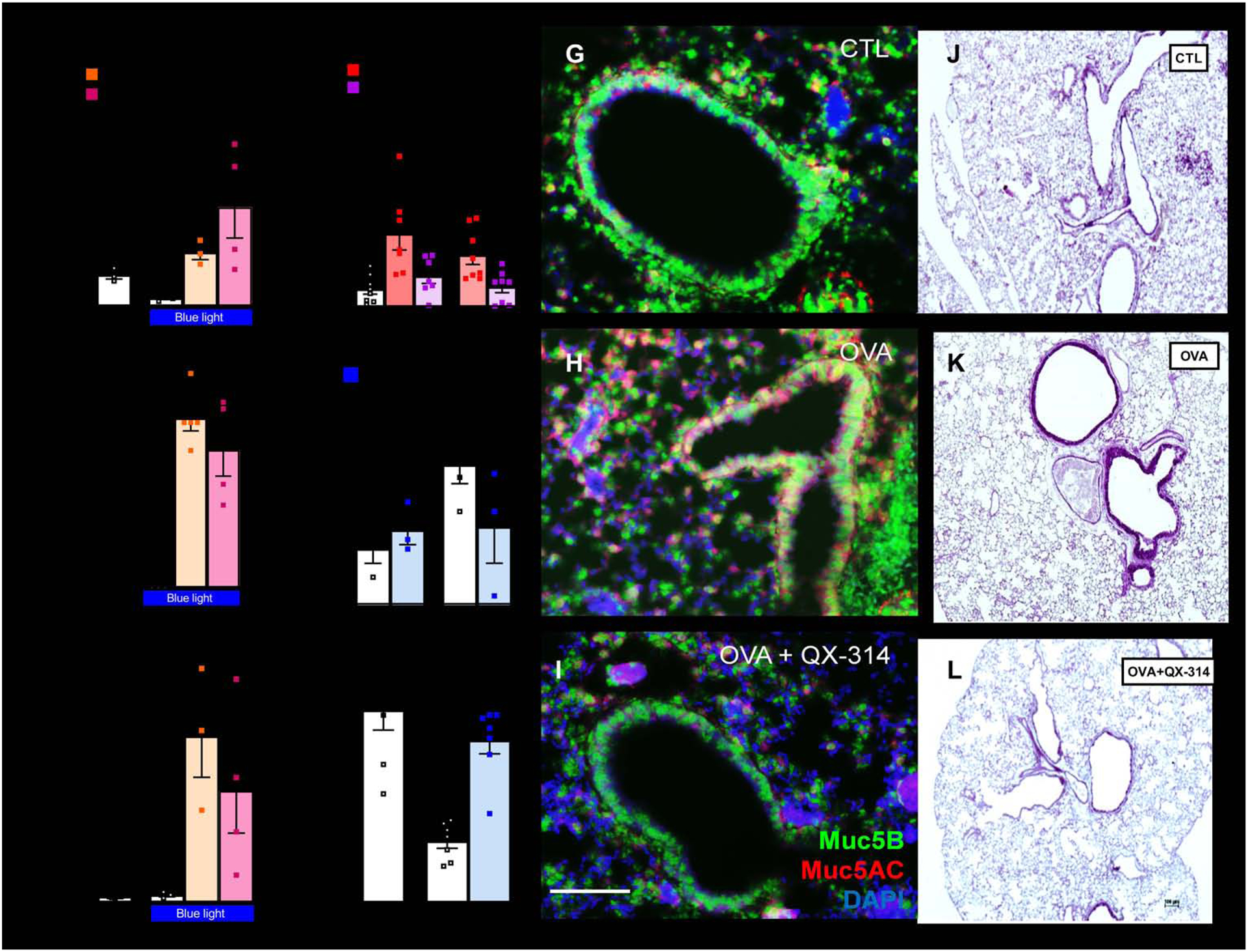
Figure 1. Airway sensory neuron neurons reverse mucus metaplasia and mucin imbalance.

Optogenetic-stimulation of TRPV1cre/wt∷ChR2fl/wt and Tac1cre/wt∷ChR2fl/wt mice vagal sensory neurons increases BALF CD45+ cells (A), lung mucus deposition (B), and BALF Muc5AC/Muc5B ratio (C). In naïve C57BL6 mice, an acute capsaicin challenge (1 uM, i.n.) increased BALF levels of Muc5AC/Muc5B. Similar findings were observed in Rag1−/− mice, suggesting that these effects are independent of B or T cells. These consequences were absent when mice were co-treated with QX-314 (100 μM, intranasal, D) to silence sensory neurons. Allergen-challenges increased airway mucus deposition (E) as well as in situ expression of Muc5AC/Muc5B (F), effects that were reversed by sensory neuron silencing using QX-314 (100 uM, aerosolized). QX-314 had no detectable outcomes when given to naïve mice (E–F). Muc5AC (red) and Muc5B (green) transcript expression (G–I) visualized by in situ-hybridization-stained sections of naïve (G) and OVA-exposed (H, I) lungs treated with saline (G, I) or QX-314 (100 μM; H). Scale 100 μm. Mucus deposition (purple; J–L) in Periodic acid–Schiff-stained sections of naïve (J) and OVA-exposed (K, L) lungs treated with saline (J, K) or QX-314 (100 μM; L). DAPI-stained cell nucleus (blue; G–I). Scale 100 μm. Mean ± S.E.M; Two-tailed unpaired Student’s t-test.
