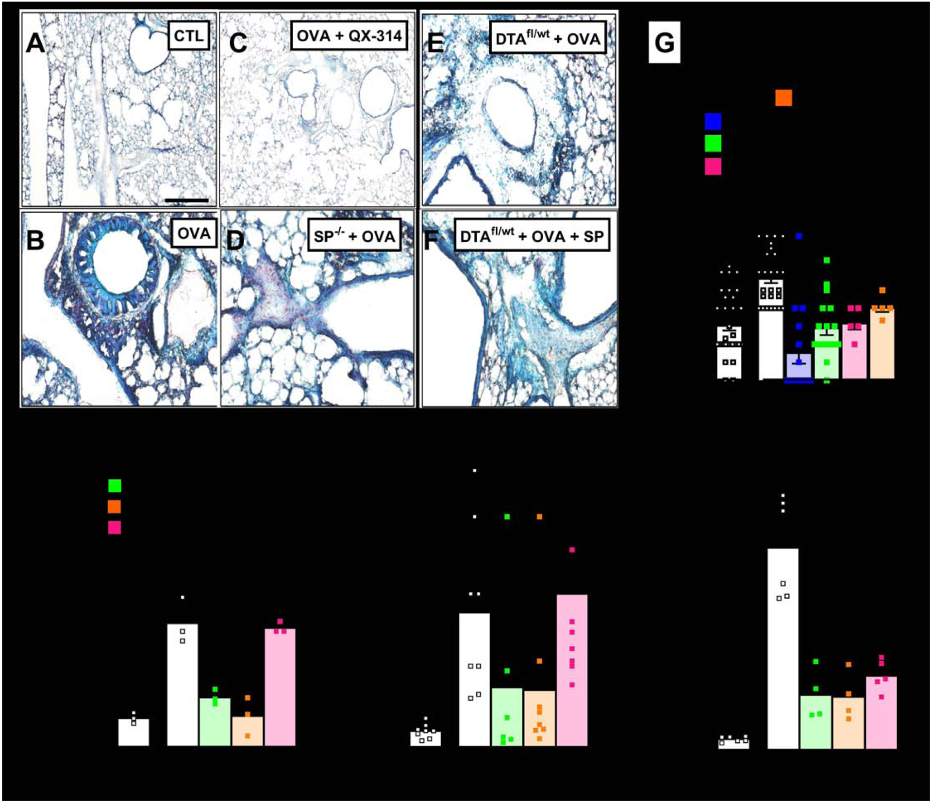
Figure 2: Allergic inflammation-mediated goblet cell hyperplasia and mucin imbalance are controlled by SP release from vagal sensory neurons.

Goblet cell hyperplasia (blue; A–F) detected in Alcian Blue (AB)-stained sections of naïve (A) and OVA-exposed (B–F) lungs from littermate control (A–C), Tac1−/− (D), or sensory neuron ablated mice (E, F) treated with vehicle (A, B, D, E), QX-314 (100 uM; C) or [Sar9, Met(O2)11]-SP (F). Scale 100 μm. Littermate control mice challenged with OVA present a significant goblet cell hyperplasia (G), mucus metaplasia (H), BALF Muc5AC/Muc5B levels (I) as well as in situ expression of Muc5AC (J) relative to naïve mice. These effects were absent in sensory neuron silenced (G), ablated (G–I) or Tac1 knockout (G–I) mice, but partially rescued by daily intranasal administration of the stable substance P analog Sar9-Met-(O2)11]-SP (G–I). Mean ± S.E.M; Two-tailed unpaired Student’s t-test.
