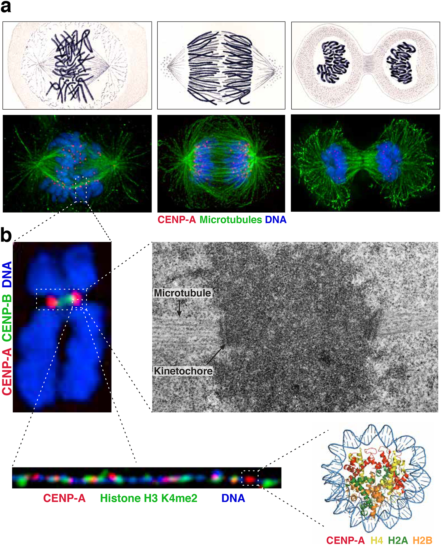
Figure 1. Visualization of the centromere.

a) Comparison of images of mitotic Salamander cells hand-drawn by Walther Flemming in 18822 (top) with immunofluorescence images of human cells (bottom) stained for microtubules (green), CENP-A (red) and DNA (blue). The images show cells at different phases of a mitotic cell cycle: late prometaphase-metaphase (left), anaphase (middle) and telophase (right). b) Images of the centromere at increasing resolution. Top left: immunofluorescence image of a mitotic chromosome stained for DNA (blue), CENP-A (red) and CENP-B (a marker for the alpha-satellite DNA repeats present at most human centromeres, green). Top right: electron micrograph of centromeric region of a mitotic chromosome showing centromeric chromatin (dark cloud), kinetochore, and microtubules (indicated by arrows). Image courtesy of Conly Rieder. Bottom left: Immunofluorescence image of stretched centromeric chromatin fibers showing patches of CENP-A (red) interspersed with H3, in this case specifically H3 dimethylated on lysine 4 (H3K4me2, green). Image courtesy of Elaine Dunleavy. Bottom right: Crystal structure of the CENP-A nucleosome90. PDB ID: 3AN2
