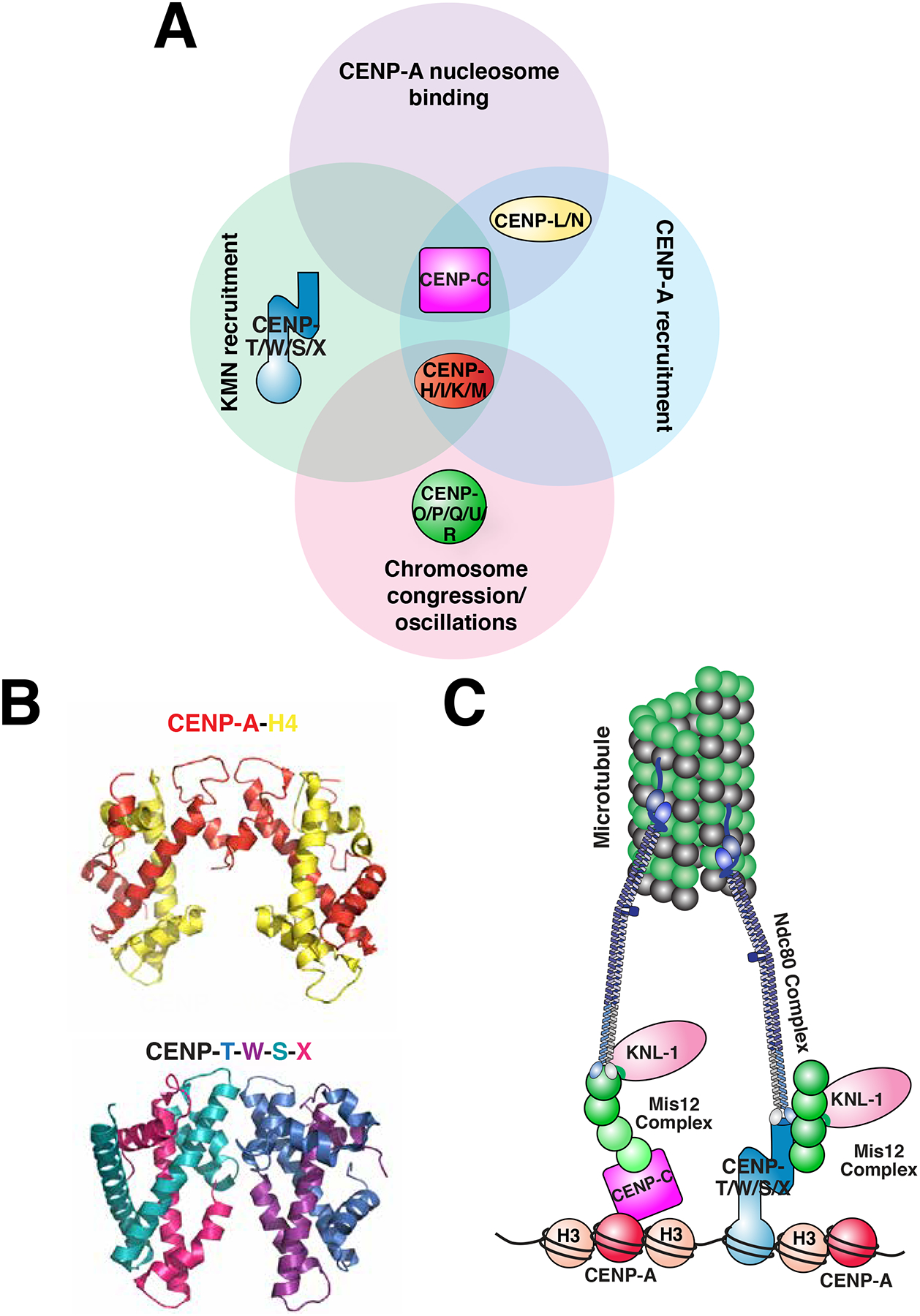
Figure 5. Contributions of the Constitutive Centromere Associated Network (CCAN) at the centromere-kinetochore interface.

a) Diagram of the proteins of the CCAN. The sixteen proteins of the CCAN, designated by CENP- and a letter, can be grouped into sub-complexes as indicated. The sub-complexes are grouped according to functions that have been reported for at least one of their subunits. KMN: a network of KNL1, Mis12 complex and Ndc80 complex, which together bind to microtubules. b) Comparison of the crystal structures of the tetramer comprised of the histones CENP-A and H4 in the context of the nucleosome (PDB ID: 3AN2)90 (H2A, H2B and DNA are excluded for clarity) with the heterotetramer comprised of the histone fold-containing proteins CENP-T, -W, -S, and -X heterotetramer (PDB 3VH5)177. c) A simplified model of the connectivity from the centromere, to the kinetochore, to the microtubule during mitosis. The contributions of CENP-C and CENP-T to recruiting the microtubule binding-interface of the kinetochore are highlighted, and the other CCAN components are excluded from this model for clarity.
