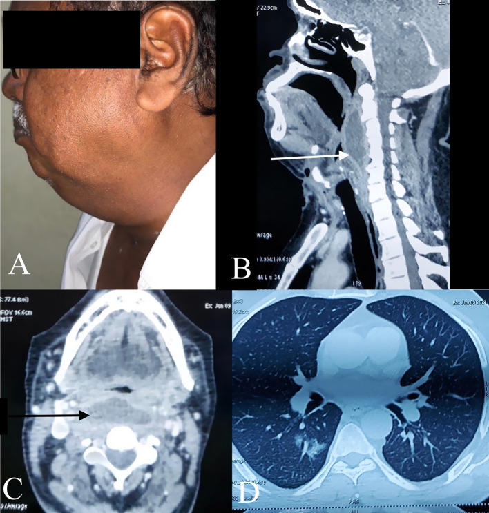
Fig. 1.
a Photograph of the patient showing the submandbibular abscess. b The computed tomography image of the neck in the lateral plane showing the retropharyngeal abscess (white arrow). c The computed tomography image of the neck in axial plane showing the retropharyngeal abscess (black arrow). d The Computed tomography image of thorax showing viral pneumonia

