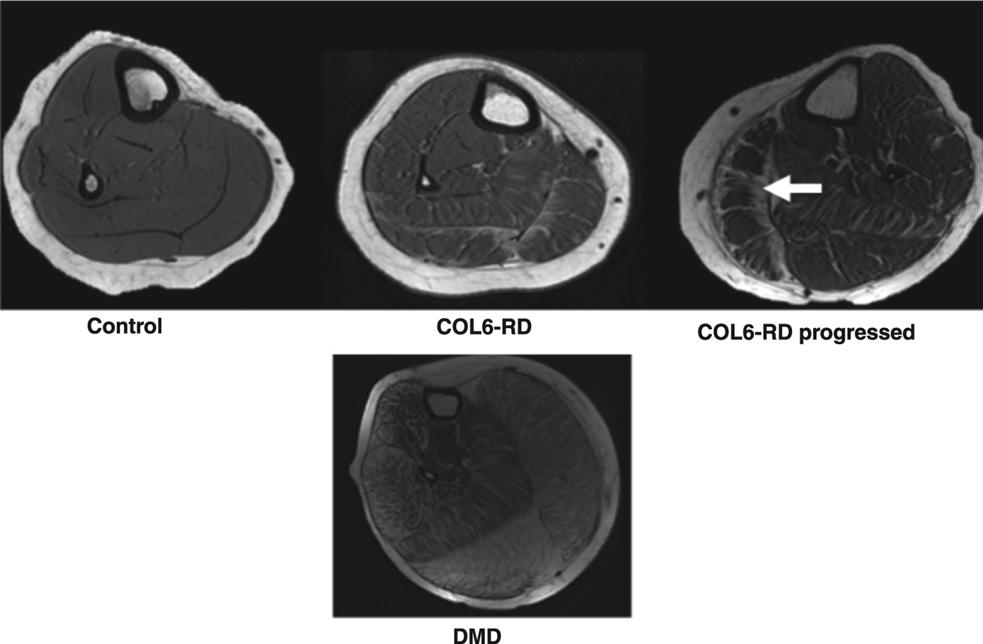
Fig. 2.

T1-weighted trans-axial images of the lower leg of age-matched patients with COL6-RD, DMD and unaffected (control) individuals showing the different patterns of disease progression with fibro-fatty infiltration in two different forms of muscular dystrophies. The characteristic inwards progression (fascia to deep inside muscle compartment) of fibro-fatty material (shown by white arrow) in the lateral gastrocnemius (LG) of subject with COL6-related dystrophy is seen. Note: Representative scans of COL6-RD progressed in figure are from left lower leg.
