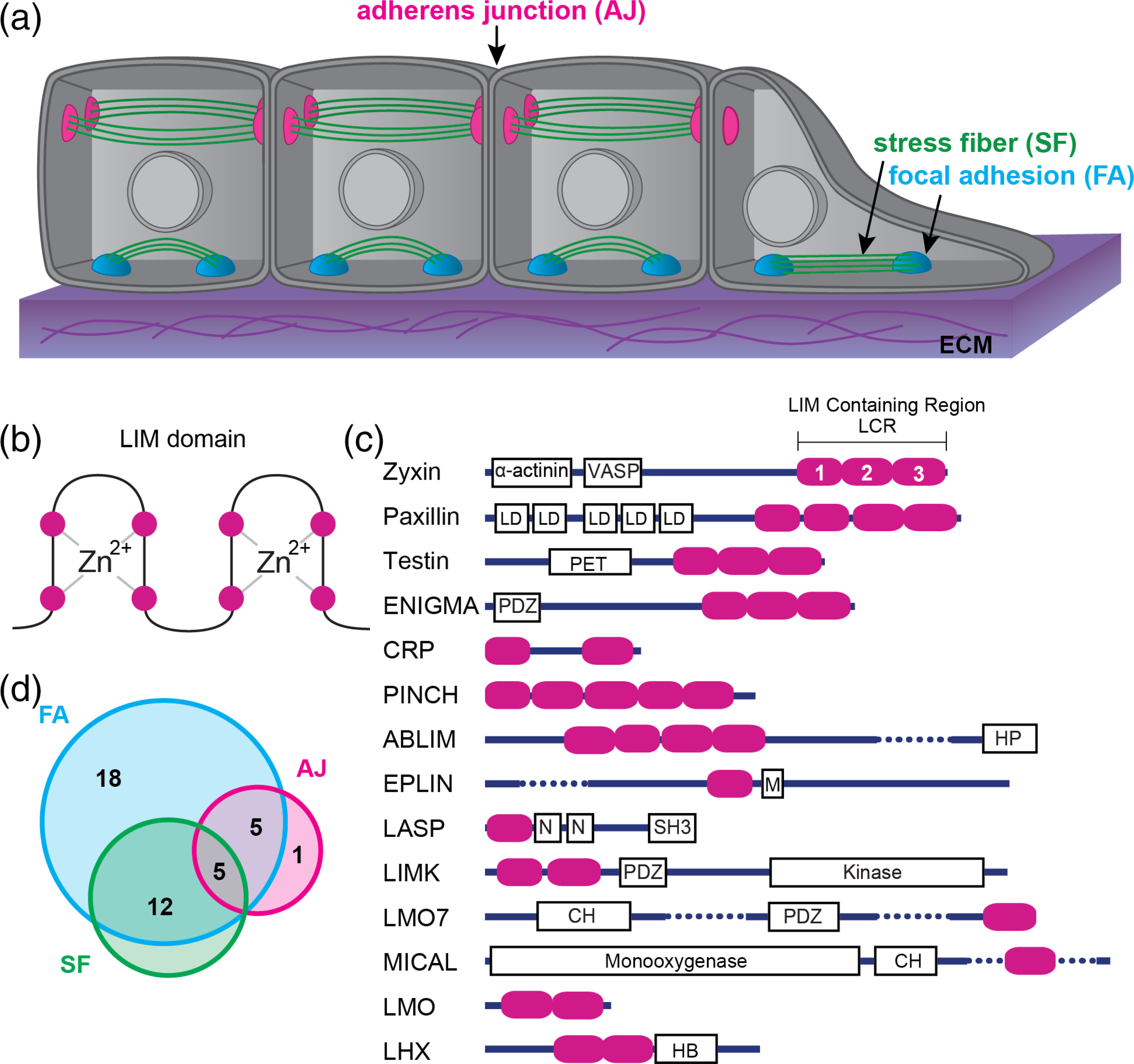
FIGURE 1.

Mechanically stressed cells and LIM domain proteins. (a) Schematic of a layer of epithelial cells on top of an extracellular matrix (ECM). (b) Simple schematic of a LIM domain: Two zinc finger motifs. The magenta circles represent the well-conserved residues (typically cysteine or histidine) that chelate the zinc molecules. The remaining amino acid sequence varies between LIM domains. (c) Domain organization of the 14 classes of LIM domain proteins. Magenta ovals represent individual LIM domains. Dotted lines are used to abbreviate a few rather long structures. Other domain abbreviations: LD, Leucine rich aspartate domains; PET, prickle, espinas, testin; PDZ, membrane anchoring domain; HP, headpiece domain for F-actin binding; M, Myo5B interacting domain; N, nebulin; SH3, Src homology 3; CH, calponin homology; HB, homeobox. (d) Venn diagram showing the overlap of LIM domain proteins that associate with the three main networks: FA, Focal adhesions; AJ, adhesion junctions; SF, stress fibers
