. 2015 Jun 11;86(2):278–284. doi: 10.2319/111714-823.1

Figure 2.

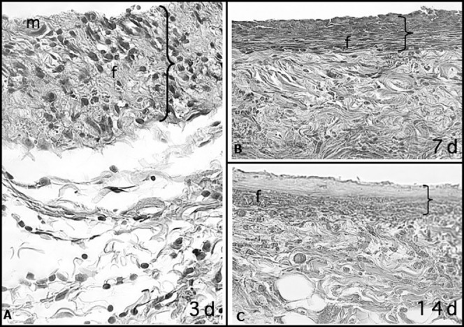
Figure 2. Capsular tissue around a nonlatex elastic with numerous macrophages (m) and few neutrophils at 3 days. At 7 and 14 days, fiber formation (f) was more intense than at 3 days and with the formation of fibrous capsule. The greater thickness of the capsule at 3 days is probably due to the greater accumulation of cells and edema, which were significantly reduced at 7 and 14 days (HE; 40× magnification).

Capsular tissue around a nonlatex elastic with numerous macrophages (m) and few neutrophils at 3 days. At 7 and 14 days, fiber formation (f) was more intense than at 3 days and with the formation of fibrous capsule. The greater thickness of the capsule at 3 days is probably due to the greater accumulation of cells and edema, which were significantly reduced at 7 and 14 days (HE; 40× magnification).