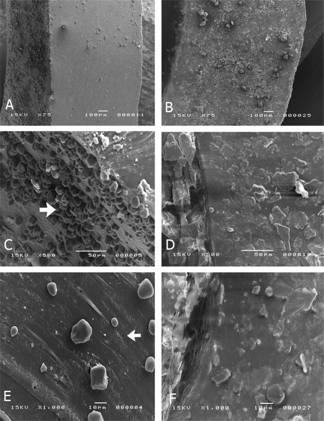
Figure 5.
SEM micrographs of latex elastics (A, C, E) and nonlatex elastics (B, D, F) at 75× to 1000× magnifications. The intact latex elastics revealed the presence of microspheres on the surface (arrow C). When sectioned, these elastics presented microspheres and porosities on their inner surface (arrow E). The intact nonlatex elastics presented crystalloid structures on the surface (B, D). When sectioned, these elastics did not present porosities (F).

